Visualizing Functional Motions of Membrane Transporters with Molecular Dynamics Simulations
By Saher A. Shaikh, Jing Li, Giray Enkavi, Po-Chao Wen, Zhijian Huang, and Emad Tajkhorshid.
Published in Biochemistry 52(4):569-87, on January 29, 2012. PMID: 23298176. PMCID: PMC3560430. Link to publication page.
Core Facility: Computational Modeling
Abstract
Computational modeling and molecular simulation techniques have become an integral part of modern molecular research. Various areas of molecular sciences continue to benefit from, indeed rely on, the unparalleled spatial and temporal resolutions offered by these technologies, to provide a more complete picture of the molecular problems at hand. Because of the continuous development of more efficient algorithms harvesting ever-expanding computational resources, and the emergence of more advanced and novel theories and methodologies, the scope of computational studies has expanded significantly over the past decade, now including much larger molecular systems and far more complex molecular phenomena. Among the various computer modeling techniques, the application of molecular dynamics (MD) simulation and related techniques has particularly drawn attention in biomolecular research, because of the ability of the method to describe the dynamical nature of the molecular systems and thereby to provide a more realistic representation, which is often needed for understanding fundamental molecular properties. The method has proven to be remarkably successful in capturing molecular events and structural transitions highly relevant to the function and/or physicochemical properties of biomolecular systems. Herein, after a brief introduction to the method of MD, we use a number of membrane transport proteins studied in our laboratory as examples to showcase the scope and applicability of the method and its power in characterizing molecular motions of various magnitudes and time scales that are involved in the function of this important class of membrane proteins.
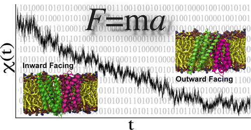
 to changes in the secondary structure and topology (middle) to changes in local interactions between individual groups at the atomic level (bottom). The structure of LeuT is used to illustrate these levels. In the top panel, LeuT (white surface) is embedded in a lipid membrane (green), surrounded by water and ions. The crystal structure is in the OF state; the cavity through which the substrate binds is visible. The middle panel shows the LeuT secondary structure, where the 10 TM helices forming the unique LeuT fold are shown. In the bottom panel, specific salt bridge and hydrophobic interactions serve as extracellular gates in LeuT. Superimposed snapshots for the residues illustrate their dynamics observed during an MD simulation.")
 Salt bridge rearrangements captured in the extracellular (EC) and intracellular (IC) halves of LeuT as the structure transits from the OF to IF state. Side chain positions before (light) and after (dark) the transition are shown. Distances between salt bridging residues show salt bridge formation as closure occurs in the EC half and breakage as opening occurs in the IC half. (B) OF to IF state transition in LeuT. Superimposed structures before (transparent) and after (colored) the transition viewed along the plane of the membrane (left) and perpendicular to the membrane: the extracellular half (right, top) and intracellular half (right, bottom). A contact map of interactions broken (black) and newly formed (red) during the transition, pinpointing conformational hot spots (green ovals).")
 Overview of the substrate release trajectory shown during equilibrium MD (pink) and SMD (red). Residues lining the substrate pathway are shown as sticks. The substrate (galactose) and the suggested gate residue, Y263, are shown in van der Waals form. (B) Time evolution of the z coordinate of the Na+ ion during six independent simulations highlighting its spontaneous unbinding from the putative site in the crystal structure. The blue bar highlights the region in the vicinity of D189. (C) Position of the substrate during a 200 ns equilibrium simulation shown using the maximal and minimal z coordinates (blue solid lines) and the geometrical center (black solid line) of the substrate. For comparison, the z position of the geometrical center of the ring of Y263 (red solid line) is also shown. Two arrows highlight the two observed substrate unbinding events from the binding site.")
 Trajectory of Pi binding (left) starting from its initial position (van der Waals representation, red) to its final binding site (blue). K80 acts as a “fishing hook”, catching the substrate at the mouth of the lumen and escorting it to the binding site at the apex of the lumen where it mainly interacts with R45 and H165. Distances between Pi and key residues in the binding site (right). No direct contact between Pi and R269 is observed during the simulations. (B) Substrate-induced helical motion in GlpT. H5 and H11 become straight upon substrate binding on the cytoplasmic side, resulting in partial occlusion of the transporter in this region. Three superimposed conformations of H5 and H11 (left): initial (t = 0; black) and final (t = 50 ns; red) along with an intermediate snapshot (t = 25 ns; blue). Distances between the H5 and H11 helices (right) measured by Cα atoms of representative residues located on the same x–y plane. All substrate binding simulations result in closure of the cytoplasmic side, while apo system simulations do not. Different substrate binding simulations are colored differently.")
 The local rearrangements of the nucleotide binding site induced by ATP hydrolysis, in particular disruption of a key hydrogen bond between the γ-phosphate and a serine residue (the thick dotted line in the left panel) at the dimer interface, eventually trigger the separation of the two monomers. The distance between the hydrogen bond donor and the acceptor in each binding site is shown at the right. (B) Global conformational change induced by ATP hydrolysis. In this particular simulation, the opening of the dimer is evident at both nucleotide binding sites. The distances between two nucleotide binding motifs at the dimer interface are recorded as an indicator of the degree of dimer opening.")
