New Software

Conformational transitions and alternating-access mechanism in the sarcoplasmic reticulum calcium pump

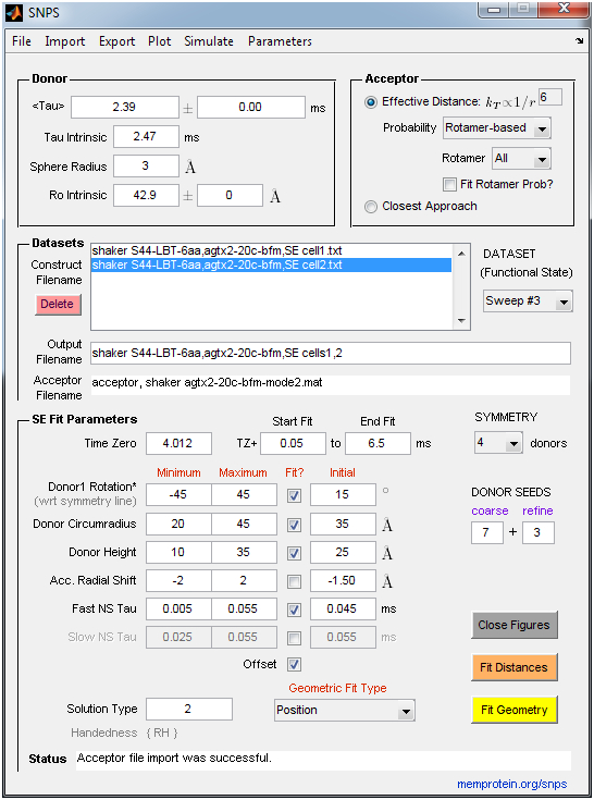
Avisek Das, Huan Rui, Lei Huang, Robert Nakamoto & Benoît Roux

http://www.sciencedirect.com/science/article/pii/S0022283617300219

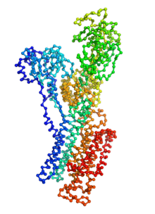
Ion pumps are integral membrane proteins responsible for transporting ions against concentration gradients across biological membranes. Sarco/endoplasmic reticulum Ca2+-ATPase (SERCA), a member of the P-type ATPases family, transports two calcium ions per hydrolyzed ATP molecule via an “alternating-access” mechanism. While X-ray crystallography provides high-resolution snapshots about the stable experimentally resolved states of the transport cycle (Figure 1), it is very difficult to detect the short-lived intermediate conformations occurring transiently during the transport cycle.

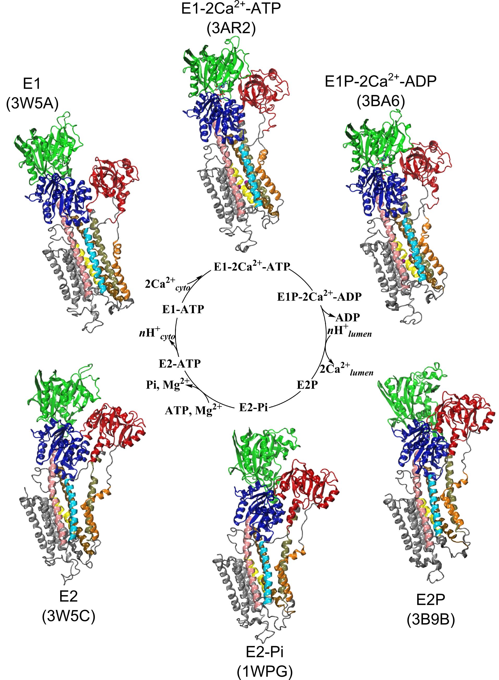
Figure 1: Structures of relevant functional states. The transport cycle of SERCA along with the structures of functional states used in the present work. The Protein Data Bank (PDB) id of each state is given in parenthesis below the state name.

Computational methods can be used to help supplement the missing information by providing atomic models for the transient intermediates along the transport cycle. Our goal with the present effort was to elucidate the details of the alternating access mechanism in SERCA by simulating the large-scale conformational transitions between the experimentally resolved stable sates. We calculated the pathway for three key steps:

  • Cytoplasmic calcium binding and occlusion (E1 to E1-2Ca2+-ATP). Under normal physiological conditions, the pump is activated by the binding of Ca2+ ions and ATP, leading to the occlusion of the pump and the hydrolysis of ATP. A complete chronology of the binding of Ca2+ ions and the concomitant large-scale conformational changes leading to the occluded state are provided by the string pathway between the E1 and the E1-2Ca2+-ATP states.
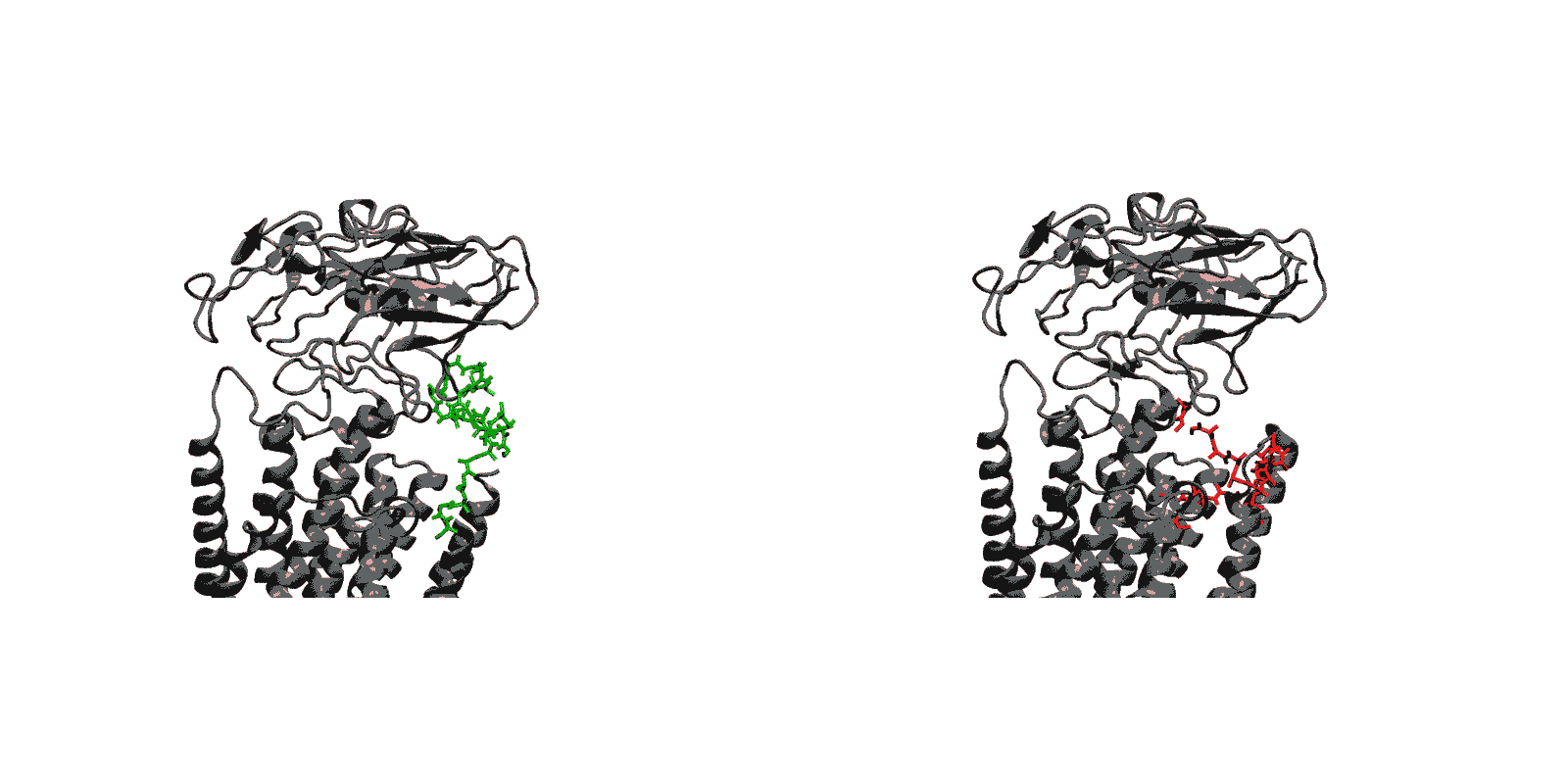
  • Luminal opening and calcium release (E1P-2Ca2+-ADP to E2P). Once the protein is stably locked in the occluded E1-2Ca2+-ATP state for an extended period of time, the phosphoryl transfer chemical reaction between ATP and Asp351 is expected to occur spontaneously, bringing the pump to the E1P-2Ca2+-ADP state (this reaction is not simulated here). This leads us to the next step in the transport cycle, which is the isomerization and conversion of the phosphorylated pump into the E2P state, where the luminal access channel is open.
  • Closing of luminal gate and dephosphorylation (E2P to E2-Pi). After the release of Ca2+ ions, the next step in the transport cycle corresponds to the transition from the E2P state to the E2-Pi state, comprising two important molecular events: the closing of the luminal gate, and the dephosphorylation of Asp351 in the P domain.

Summary

The computational pathways provide a clear chronology of the key events underlying the active transport of calcium ions by SERCA. The main conclusions regarding three critical steps of the transport cycle E1→E1-2Ca-ATP→E2P→E2-Pi are:

  • (E1 to E1-2Ca-ATP) Starting from the cytoplasmic open-access E1 state, the binding of ATP and the formation of the phosphoryl transfer catalytic site at the interface of the N and P domain together with the binding and sequestration of the two Ca2+ ions in the TM binding sites leading to occlusion are highly concerted and cooperative. The highly concerted transition explains the tight coupling between Ca2+ binding and ATP hydrolysis that is observed experimentally.
  • (E1-2Ca-ATP to E2P) Phosphorylation of Asp351 when the pump is in the occluded E1-2Ca-ATP state drives the opening of the luminal gate via a two-step mechanism. In a first step, phosphorylation of Asp351 triggers a large-scale rotational movement of the cytoplasmic A domain to form a strong interaction with Asp351-P at the surface of the P domain (blocking of the catalytic site and preventing the back reaction by the ADP). In a second step, the large movement of the A domain is subsequently transmitted to the M4 helix via M3 helix leading to the disruption of the Ca2+ binding sites and the opening of the TM region toward the luminal side.
  • (E2P to E2-Pi) The closing of the luminal gate following the dissociation of the Ca2+ ions to the luminal solution and protonation of binding sites residues is correlated with the dephosphorylation of Asp351-P via a two-step mechanism. In a first step, dehydration of the (protonated) binding sites together with the hydrophobic re-packing between the TM helices M4, M5 and M6 spontaneously drives the closing of the luminal gate. In a second step, the closed luminal gate mediated by the junction with the M2 helix induces a rotation in the position of the A domain, positioning the 181TGES loop in the A domain together with increased water access near Asp351-P needed for the dephosphorylation reaction.

Acknowledgments

This work was supported by the Membrane Protein Structural Dynamics Consortium funded by NIH/NIGMS through grant U54-GM087519. An award of computer time was provided by the Innovative and Novel Computational Impact on Theory and Experiment (INCITE) program from the Department of Energy (DOE) of the United States of America. This research used resources of the Argonne Leadership Computing Facility (ALCF), which is a DOE Office of Science User Facility supported under Contract DE-AC02-06CH11357. Additional computational resources were provided by the Great Lake Consortium for Petascale Computing and the XSEDE program of the National Science Foundation of the United States of America.

New Webserver: ANMPathway determines the most probable (lowest energy) transition pathway between two stable endpoints of a conformational transition

Biomolecular conformational transitions are essential to biological functions. Most experimental methods report on the long-lived functional states of biomolecules, but information about the transition pathways between these stable states is generally scarce. Such transitions involve short-lived conformational states that are difficult to detect experimentally. For this reason, computational methods are needed to produce plausible hypothetical transition pathways that can then be probed experimentally. Here we propose a simple and computationally efficient method, called ANMPathway, for constructing a physically reasonable pathway between two endpoints of a conformational transition. We adopt a coarse-grained representation of the protein and construct a two-state potential by combining two elastic network models (ENMs) representative of the experimental structures resolved for the endpoints.

ANMPathway determines the most probable (lowest energy) transition pathway between two stable endpoints of a conformational transition. Conceptually, ANMPathway represents a direct application of the string method to a two-state CG system approximated by ANM energy surfaces. The result is a sequence of PDB structure (a “string”) smoothly linking the two endpoints.

Goals

  • Generates a continuous minimum energy pathway from two End-Point PDB structures containing the same number and order of residues.
  • ANMPathway sends the calculated pathway by email. Typical jobs take several minutes to an hour (maximum 12 hours). The result also contains a movie of the structural transition along the calculated pathway and a list of non-native contacts formed during the transition.
  • NAMD scripts for converting the CG pathway to an all-atom pathway is coming soon.

Webserver: http://anmpathway.lcrc.anl.gov/anmpathway.cgi/

This webserver was developed by Avisek Das and Sunhwan Jo from Benoit Roux’s group at the University of Chicago. Please contact us with questions or suggestions using the comments form below.

New webserver: prediction of LBT (lanthanide-binding tag) insertion onto membrane proteins

Background

In recent years, high-throughput genomic and proteomic projects have been revealing new genetic information at a very fast pace. The proper interpretation of such information in the context of the underlying biological mechanisms generally requires studies on a molecular level through biophysical and biochemical experiments as well as biomolecular modeling. Hence, the use of biophysical probes that can be easily integrated into protein structures for functional and structural characterization is becoming increasingly common. Lanthanide-binding tags (LBTs) are a class of such probes that are optimized for binding lanthanide ions.

We have created a fully automated webserver for the prediction of LBT insertions onto existing protein structures. The underlying method uses a coarse-grained backbone+Cβ representation of the protein and samples on the backbone dihedral angles. The method predicts the proper conformation of the LBT tag with respect to the parent protein in existing fusion crystal structures. On multiple membrane protein systems, the method’s prediction of feasibility of LBT insertion on various sites qualitatively agree with the experimental data. The method currently reports several metrics to assess the quality of LBT insertion in a specific site in a parent protein. We expect the method to serve as a useful computational tool for experimentalists by helping them select reasonable sites in a given parent protein where the LBT insertion will likely be successful.

Goals

  1. Given a parent protein where the LBT tag is to be inserted, the method will predict the likelihood of a successful LBT insertion at a certain site in the parent protein.
  2. Given a parent protein and a site of insertion, the method will predict the proper conformation of the parent+LBT fusion conformation.

Webserver

http://goo.gl/1m0Yib

This webserver was developed by Aashish Adhikari from Tobin Sosnick’s group at the University of Chicago. Please contact us with questions or suggestions using the comments form below.

GAAMP (General Automated Atomic Model Parameterization) XSEDE Gateway made available

We are happy to announce that the Computational Modeling Core has made an XSEDE gateway available for its GAAMP: General Automated Atomic Model Parameterization force field server.

All-atom force fields are mathematical objects constructed from simple analytical functions parameterized to approximate the Born-Oppenheimer potential energy surface and reproduce known experimental observables. Parameters for the all-atom additive non-polarizable potential functions are currently available for amino acids, nucleic acids and common phospholipids. But accurate potential functions are also required for a growing number of novel molecules. Benoît Roux’s group at the University of Chicago has developed this Force Field Server for automatically generating testing and validating the all-atom nonpolarizable force fields used in MD simulations based on quantum mechanical (QM) calculations.

This GAAMP gateway is developed to help XSEDE users in using GAAMP for automated force field parameterization, based on the power of XSEDE computing resources. Currently, the trestles.sdsc.edu interface is being utilized. An interface for Blacklight users will be ready soon as well.

Visit the GAAMP XSEDE Gateway »

New SNPS (Symmetric Nano-Positioning System) software made available

Proteins may undergo multiple conformational changes required for their function. One strategy used to estimate target site positions in unknown structural conformations involves single-pair resonance energy transfer (RET) distance measurements. However, interpretation of inter-residue distances is difficult when applied to 3D structural rearrangements, especially in homomeric systems. Probe diffusion further complicates this task by biasing measurements towards shorter distances. Lanthanide resonance energy transfer (LRET) is an ideal technique for simultaneously resolving multiple distances within a protein. Hyde et al. (2012) recently combined LRET with an ensemble of numerical methods to form the Symmetric Nano-Positioning System (SNPS), which allows accurate 3D positioning and inter-probe distance estimation in functional homomeric proteins.

SNPS determines the 3D position of lanthanide donors [satellites] attached to a target site (one per subunit), relative to a single fluorescent acceptor [antenna] placed in a static reference site, as illustrated in the above figure. The acceptor’s position and accessible volume can be modeled from its structure-based labeling site by several methods, including dihedral scan analysis. SNPS can be applied to all defined conformational states of the protein and with simultaneous functional recordings. Satellite-to-antenna distances are encoded in time-resolved LRET lifetime decays. SNPS directly fits a 3D geometric model of satellite positions to LRET lifetime measurements using an inverse trilateration-based curve fitting procedure. Global analysis implementation fits the geometric model to an ensemble of replicate measurements. Numerical and analytical tools are integrated to account for probe diffusion and evaluate the confidence region of fitted positions. SNPS is well-suited to estimate 3D conformational changes at the target site between defined conformational states. In its first application, SNPS was used to determine the position of a functional voltage-gated potassium channel’s voltage sensor in its three major conformations [H. Clark Hyde, Walter Sandtner, Ernesto Vargas, Alper T. Dagcan, Janice L. Robertson, Benoit Roux, Ana M. Correa, Francisco Bezanilla (2012), Structure 20(10): 1629-1640].

The SNPS Toolbox contains the following stand-alone programs that implement the SNPS method:

  • SNPS: Performs inverse trilateration-based curve fitting of LRET lifetime decays to estimate the 3D position of lanthanide donors [satellites] attached to a target site (one per subunit), relative to a single fluorescent acceptor [antenna] placed in a static reference site. An acceptor file must be supplied by the user that specifies the acceptor’s coordinates and accessible volume. SNPS can alternatively fit all donor-acceptor distances constrained such that donors must lie in a polygon (symmetric) geometry, without any knowledge of acceptor position. SNPS can also simulate LRET lifetime decays at a user-defined donor position and noise level to explore the relation between donor position and decay shape. LRET lifetime decays are expected to be sensitized emission measured by a photomultiplier tube in analog mode (Poisson noise). The user imports lifetime decay files (ASCII .txt) and an acceptor file (ASCII .txt or Matlab .mat). See the “Example Data” folder for examples of required formatting. Fits are accompanied by a comprehensive set of output figures and a solution file.
  • DecayAnalysis: Performs model-free curve fitting of multi-exponential decays to estimate time constants and amplitudes (up to 4 exponential components). It is intended for donor-only time constant analysis of LRET experiments, but can also be used for general exponential fitting. The donor-only time constant is a required parameter for the SNPS program. The user can place constraints on time constants and define a weights scheme appropriate for the measurement uncertainty (e.g., Poisson vs. Gaussian noise). A large set of measurements can be fit quickly with basic statistical analysis reported. The user imports lifetime decay files in ASCII (.txt) format. See the “Example Data” folder for examples of required formatting.

SNPS and Decay Analysis Screen Guides


Download the software (version 2012.1):
Windows 64-bit: SNPS_Toolbox_win64.exe
Windows 32-bit: SNPS_Toolbox_win32.exe

Installation: Clicking on the downloadable .exe file will directly launch the Matlab Component Runtime (MCR) installation process and extract the SNPS and DecayAnalysis applications. This is a one-time installation, after which both applications can be launched. Installation of the included MCR is required even if you currently have Matlab or other MCR versions installed. Regarding processing speed, CPU speed takes precedence over the number of cores. The extracted folder also contains an “Example Data” folder with example data from the publication: AgTx2[II]-D20C-BODIPY-FL-maleimide acceptor cloud and Shaker S4(4) LBT construct sensitized emission (SE) and donor-only (DO) lifetime decays. Fit progress and information is displayed in an accompanying DOS window. Output files are written to a subfolder of the imported data directory.

The software was developed and written by H. Clark Hyde from Francisco Bezanilla’s group at the University of Chicago. Please contact us with questions or suggestions using the comments form below.

New Continuum-Molecular Dynamics (CTMD) software made available by Computational Modeling Core

The membrane environment is a key determinant of function and/or organization of membrane proteins. It is essential for the activities of the Consortium to have access to quantitative information about the characteristics and energetics of membrane-protein interactions. For multi-segment transmembrane proteins in particular, such characterization is complicated by the different hydrophobic thicknesses of the component transmembrane segments and the radially non-uniform hydrophobic interface created between the membrane and the protein. A hybrid Continuum-Molecular Dynamics (CTMD) approach to compute membrane deformations profiles around multi-segment proteins and corresponding energetics was recently published [Sayan Mondal, George Khelashvili, Jufang Shan, Olaf Andersen, Harel Weinstein (2011), Biophysical J (101): 2092-2101; link]. This stand-alone application implements the CTMD approach.

Protocol for the 3D-CTMD approach, illustrated for rhodopsin in a diC14:1PC lipid bilayer (Mondal et al., BJ 2011). Click to enlarge and learn more.

The CTMDapp software calculates the deformation profiles of the bilayer and the free energy cost of the membrane deformation around multi-segment transmembrane proteins, taking into account the radially non-uniform hydrophobic surface of the protein. As the primary input it requires a molecular dynamics trajectory of a multi-segment, transmembrane protein embedded in a bilayer.

To allow for calculations without a molecular dynamics input, the CTMDapp software also implements a simplified version of the CTMD method that can assess the radial asymmetry of the membrane-protein interface for a particular protein structure at an approximate level.

Methodological details can be found in the original paper (Mondal et al., BJ 2011). In addition, the application is documented with brief usage notes at every step and generates diagnostic intermediate output.

Download the software:

Instructions:
For the Windows version, clicking on the downloadable .exe file will directly launch the Matlab Component Runtime (MCR) installation process and extract the app executable. For the Mac version, the MCR must be installed first by opening MCRinstaller.dmg in the folder. This is a one-time installation, after which CTMDapp can be launched. The Unix version requires an interface like Xwin along with a script setting proper paths.

The software was written by Sayan Mondal (mos2013@med.cornell.edu) in the Harel Weinstein (haw2002@med.cornell.edu) lab at Weill Cornell Medical College, Cornell University. Please feel free to contact us with questions and suggestions about the software.

Categories
Recent Posts
Archives
Recent Comments
Recent Photos