Advances in the Field

Active MPSDC participation in the Biophysical Society 60th Annual Meeting in 2016


MPSDC Awards: Francisco Bezanilla, Past President presenting an award to Robert Nakamoto.
(Click to enlarge)

The Membrane Protein Structural Dynamics Consortium is always very well represented at the annual Biophysical Society meeting, as can be seen by Klaus Schulten‘s National Lecture in 2015 as well as at the level of participants in a remarkably large number of workshops, symposia, and presentations. This is also reflected in Consortium members being actively involved at the leadership level, including Francisco Bezanilla‘s tenure as Biophysical Society President from 2012 to 2014 and Robert Nakamoto and Olga Boudker‘s service in the current Biophysical Society leadership council.

On August 31, 2015 the Biophysical Society announced that MPSDC Chair Eduardo Perozo was elected as a 2016 Society Fellow. This award honors the Society’s distinguished members who have demonstrated excellence in science, contributed to the expansion of the field of biophysics, and supported the Biophysical Society. The Fellows will be honored at the Awards Ceremony during the Biophysical Society’s 60th Annual Meeting on Monday February 29, 2016 at the Los Angeles Convention Center in Los Angeles, California. Perozo was elected for his leadership and fundamental contributions in ion channel biophysics.

At next year’s meeting in Los Angeles, MPSDC members will participate in a number of specialty symposia and workshops organized by the Biophysical society (more information on the nature of these symposia and workshops can be found on the Biophysical Society meeting website here). We would like to highlight the following in particular (though there are and will certainly be more ways in which Consortium members are involved with the meeting):

  • Francisco Bezanilla (University of Chicago) is participating in a symposium on Voltage Sensing and Gating.
  • Olga Boudker (Weill Cornell Medical College) is receiving the Michael and Kate Bárány Award during the meeting’s award symposium. During this session, award recipients are recognized and each give a short talk about the work for which they are being recognized. Congratulations, Olga!
  • Yifan Cheng (University of California, San Francisco) is co-chairing the Cryo-EM Subgroup 2016 Symposium
  • Claudio Grosman(UIUC) is participating in a symposium on Pentameric Ligand-gated Ion Channels: New Insights from Structure and Function.
  • Benoît Roux (University of Chicago) is participating in a workshop about Computational Methods for Ion Permeation and Selectivity
  • Emad Tajkhorshid (UIUC) will once again be chairing the Permeation & Transport Subgroup 2015 Symposium. He chaired the same subgroup last year.

Last year, MPSDC member and one of our keynote speakers at Frontiers in Membrane Protein Structural Dynamics 2014 Klaus Schulten (UIUC) gave the prestigious National Lecture. The National Lecturer is the highest award given each year by the Biophysical Society. Dr. Schulten’s 2015 National Lecture lecture can be viewed in full here.

See also: interviews from the 59th Biophysical Society Meeting with 2015 National Lecturer Klaus Schulten and Harel Weinstein.

See also: interviews from the 58th Biophysical Society Meeting with Francisco Bezanilla, Past President and Robert Nakamoto, Chair of the 2014 BPS Program Committee.

Solving a Major Piece of a Cellular Mystery about Nuclear Pore Complexes

Drs. Shohei Koide and Anthony Kossiakoff recently worked together with a team to determines the architecture of a second subcomplex of the nuclear pore complex, in so doing solving a major quandary of answering how a nuclear pore complex (NPC) can be such an effective gatekeeper, preventing much from entering the nucleus while helping to shuttle certain molecules across the nuclear envelope.

The scientific team was featured in multiple publications including Caltech and the Argonne National Laboratory Science Highlights (reproduced in part below). The research was supported in part by the Consortium, and culminated in the following publication:

Architecture of the fungal nuclear pore inner ring complex

Stuwe T, Bley CJ, Thierbach K, Petrovic S, Schilbach S, Mayo DJ, Perriches T, Rundlet EJ, Jeon YE, Collins LN, Huber FM, Lin DH, Paduch M, Koide A, Lu V, Fischer J, Hurt E, Koide S, Kossiakoff AA, Hoelz A. Science. 2015 Oct 2;350(6256):56-64. PMID: 26316600.

Learn more »

Solving a Major Piece of a Cellular Mystery

This article by Argonne reproduced in part can be read here

Not just anything is allowed to enter the nucleus, the heart of eukaryotic cells where, among other things, genetic information is stored. A double membrane, called the “nuclear envelope,” serves as a wall, protecting the contents of the nucleus. Any molecules trying to enter or exit the nucleus must do so via a cellular gatekeeper known as the nuclear pore complex (NPC), or pore, which exists within the envelope.

How can the NPC be such an effective gatekeeper — preventing much from entering the nucleus while helping to shuttle certain molecules across the nuclear envelope? Scientists have been trying to figure that out for decades, at least in part because the NPC is targeted by a number of diseases, including some aggressive forms of leukemia and nervous system disorders such as a hereditary form of Lou Gehrig’s disease. Now a team of researchers from Caltech, The University of Chicago, and the Biochemistry Center of Heidelberg University (Germany), led by André Hoelz and working at three U.S. Department of Energy synchrotron light sources including the Advanced Photon Source (APS) at Argonne, has solved a crucial piece of the puzzle.

In February of this 2015, the team published a paper describing the atomic structure of the NPC’s coat nucleoporin complex, a subcomplex that forms what they now call the outer rings (see the figure). Building on that work, the team has now solved the architecture of the pore’s inner ring, a subcomplex that is central to the NPC’s ability to serve as a barrier and transport facilitator. In order to determine that architecture, which determines how the ring’s proteins interact with each other, the biochemists built up the complex in a test tube and then systematically dissected it to understand the individual interactions between components. Then they validated that this is actually how it works in vivo, in a species of fungus.

Read more »

Researchers discover structure of fluoride-specific ion channel

Dr. Shohei Koide‘s collaborations in a scientific team seeking to discover the structure of a fluoride-specific ion channel was recently featured in Brandeis NOW, a scientific research publication published by Brandeis University. The work focuses on Dr. Christopher Miller’s lab, based at Brandeis University. The research was supported in part by the Consortium, and culminated in the following publication:

Crystal structures of a double-barrelled fluoride ion channel

Stockbridge RB, Kolmakova-Partensky L, Shane T, Koide A, Koide S, Miller C, Newstead S. Nature. 2015 Sep 24;525(7570):548-51. PMID: 26344196.

Learn more »

Researchers discover structure of fluoride-specific ion channel

Creation of ‘atomic blueprint’ is a biological novelty

The original Brandeis NOW press release by Kimm Fesenmaier can be read here

Fluoride protects our teeth against cavity-causing bacteria by making our teeth stronger. But what if we could find a way to trap fluoride ions (the negatively charged form of the chemical element fluorine) inside bacteria? At the right concentration, fluoride ions are highly toxic to bacteria, wreaking havoc on their proteins and disrupting critical cellular functions. Bacteria, however, can fight back, exporting the toxic fluoride ions out using specialized proteins called fluoride-specific ion channels.

How these proteins remove fluoride ions from the cell is poorly understand. To glimpse into the inner workings of these proteins, researchers in Christopher Miller’s lab at Brandeis University in collaboration with Simon Newstead at the University of Oxford have determined the structure of one such fluoride-specific ion channel from the Bordetella pertussis bacteria called Bpe.

In a paper published by Nature on Sept. 7, lead author Randy Stockbridge, a post-doctorate fellow in Miller’s lab, and colleagues used a technique called X-ray crystallography to obtain an “atomic blueprint” illustrating the arrangement of the amino acids that make up Bpe. The details provide important insight into how Bpe exports fluoride out of the cell. Intriguingly, the schematics also point out potential weaknesses in Bpe that could be exploited to trap fluoride inside bacteria.

Based on the blueprint, the Bpe fluoride-specific ion channel resembles an hourglass and is actually composed of two Bpe protein molecules. At the center where the hourglass constricts is a sodium ion that may act like a pin that fastens the two Bpe proteins together. However, rather than having a central pore like an hourglass, the arrangement of the two Bpe molecules forms two parallel tunnels through which fluoride ions could flow. The blueprint also helps explain why Bpe only exports fluoride; the tunnels are just the right size for fluoride ions, but are too narrow for the biologically abundant chloride ion, fluoride’s larger but chemically similar cousin.

Notably, the researchers observed two unidentifiable “hazy shadows” in each tunnel, which they concluded were fluoride ions. First, many of the amino acids along the walls of each channel that protrude toward the shadows are chemically attracted to fluoride ions. Consistently, when the researchers mutated some of these amino acids to change their chemical properties, Bpe’s ability to export fluoride ions out of the bacteria was dramatically reduced. The researchers also studied the structure of a related fluoride-specific ion channel, Ec2, and found similar hazy shadows in its tunnels.

The researchers also noted a peculiar feature of Bpe that has implications for how the protein moves fluoride out. At one point in each of the tunnels, the side chain, or chemical appendage, of a particular amino acid protrudes inward, contacts the fluoride ion, and impedes its path to the exit. The side chain can swivel though, so it may be that the fluoride ion grasps it as though it were a turnstile and is then pulled to the other side when the side chain rotates.

Armed with Bpe’s blueprints, researchers can exploit structural weaknesses and develop strategies that kill bacteria by preventing fluoride from being moved out. Chemical compounds could be designed that pull the sodium ion pin and dismantle Bpe or that lock the turnstile causing a fluoride traffic jam that backs up into the cell.

In addition to Stockbridge, Miller and Newstead, the paper’s other authors are Ludmila Kolmakova-Partensky, Tania Shane, Akiko Koide and Shohei Koide.

The research was supported in part by a Wellcome Trust Investigator Award and grants from the National Institutes of Health (NIH) (RO1-GM107023 and U54-GM087519). Stockbridge also was supported by an NIH grant (K99-GM-111767). Miller is a Howard Hughes Medical Investigator.

Highlights and overview of selected recent Consortium-sponsored publications

Biomolecular DNP- supported NMR spectroscopy using site directed spin labeling

Authors: Elwin A.W. van der Cruijsen, Eline J. Koers, Claire Sauvee, Raymond E. Hulse, Markus Weingarth, Olivier Ouari, Eduardo Perozo, Paul Tordo, and Marc Baldus.

In the press, Journal of the American Chemical Society. (Consortium project: Dynamics of Ion Permeation and Conformation Coupling in KcsA)

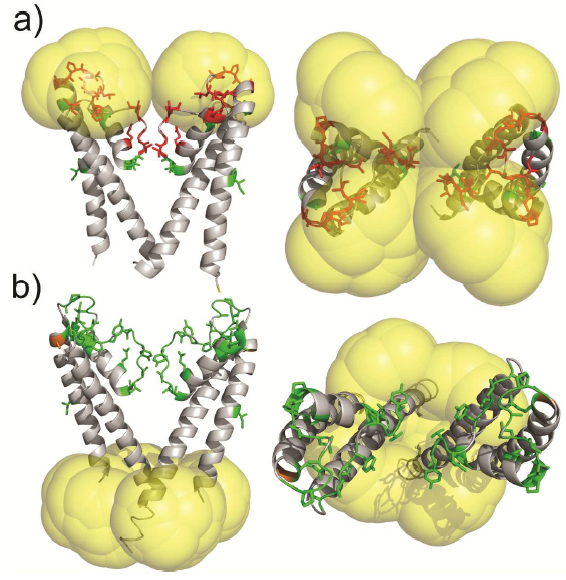
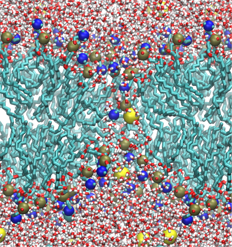
Molecular probes that combine the benefits of enhanced spectroscopic sensitivity with site-specific localization have significantly expanded our ability to track molecular structure and function in applications ranging from cell biology to material science . In the field of magnetic resonance, dynamic nuclear polarization (DNP) has become a widely usable method to significantly enhance overall spectroscopic sensitivity in NMR and MRI. Here, the authors show that DNP can be established by creating local spin clusters via site-directed spin labeling using mono- or biradicals. Applied to a membrane-embedded potassium channel, we show that this approach can significantly enhance NMR sensitivity while preserving the intrinsic spectroscopic properties of (bi)radicals as paramagnetic relaxation enhancers.

The results suggest that the creation of local spin clusters can generate sizable DNP enhancements while preserving the intrinsic benefits of PRE-based NMR approaches. Our results are consistent with the idea that the magnitude in DNP enhancement are highly dependent on the nearest neighbor electron-electron distances.

13C NMR detects conformational change in the 100-kD membrane transporter Cl-C-ec1

Authors: Sherwin J. Abraham, Ricky C. Cheng, Thomas A. Chew, Chandra M. Khantwal, Corey W. Liu, Shimei Gong, Robert K. Nakamoto, and Merritt Maduke.

J Biomol NMR. 2015 Jan 29. (Consortium project: Conformational Dynamics in the CLC Channel Transporter Family. Consortium core: Membrane Protein Production)

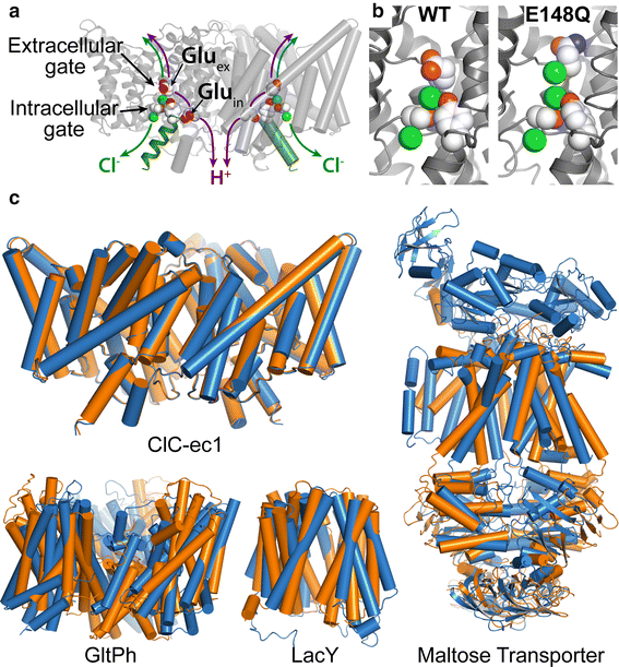
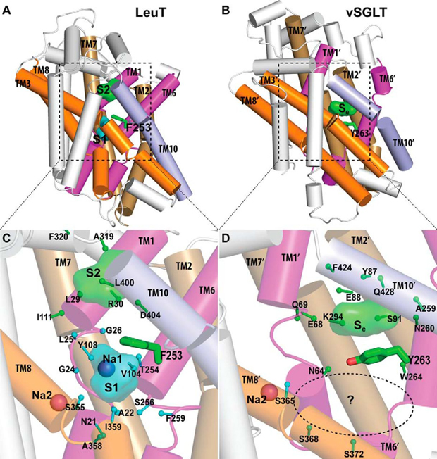
Members of the Cl-C (‘‘Chloride-Channel’’) family play central roles in cardiovascular, neuronal, bone, and epithelial function. Cl-C transporters catalyze the exchange of Cl--for H+ across cellular membranes. To do so, they must couple Cl-- and H+ binding and unbinding to protein conformational change. However, the sole conformational changes distinguished crystallographically are small movements of a glutamate side chain that locally gates the ion transport pathways. Therefore, our understanding of whether and how global protein dynamics contribute to the exchange mechanism has been severely limited. To overcome the limitations of crystallography, the authors used solution-state 13Cmethyl NMR with labels on methionine, lysine, and engineered cysteine residues to investigate substrate (H+) dependent conformational change outside the restraints of crystallization. They show that methyl labels in several regions report H+-dependent spectral changes. They  identify one of these regions as Helix R, a helix that extends from the center of the protein, where it forms the part of the inner gate to the Cl-–permeation pathway, to the extracellular solution. The H+-dependent spectral change does not occur when a label is positioned just beyond Helix R, on the unstructured C-terminus of the protein.

Together, the results suggest that H+ binding is mechanistically coupled to Cl-osing of the intracellular access-pathway for Cl--. These studies set the stage for investigating the structural details and dynamics of this change.

Room-Temperature Distance Measurements of Immobilized Spin-Labeled Protein by DEER/PELDOR

Authors: Virginia Meyer, Michael A. Swanson, Laura J. Clouston, Przemyslaw J. Boratynski, Richard A. Stein, Hassane S. Mchaourab, Andrzej Rajca, Sandra S. Eaton, and Gareth R. Eaton.

Biophys J. 2015 Mar 10;108(5):1213-9. (Consortium Core: Membrane Protein Production)

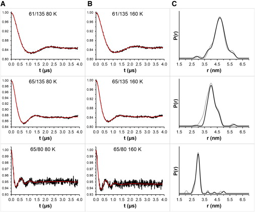
Nitroxide spin labels are used for double electron-electron resonance (DEER) measurements of distances between sites in biomolecules. Rotation of gem-dimethyls in commonly used nitroxides causes spin echo dephasing times (Tm) to be too short to perform DEER measurements at temperatures between ~80 and 295 K, even in immobilized samples. A spirocyclohexyl spin label has been prepared that has longer Tm between 80 and 295 K in immobilized samples than conventional labels. Two of the spirocyclohexyl labels were attached to sites on T4 lysozyme introduced by site-directed spin labeling. Interspin distances up to ~4 nm were measured by DEER at temperatures up to 160 K in water/glycerol glasses. In a glassy trehalose matrix the Tm for the doubly labeled T4 lysozyme was long enough to measure an interspin distance of 3.2 nm at 295 K, which could not be measured for the same protein labeled with the conventional 1-oxyl-2,2,5,5-tetramethyl-3-pyrroline-3-(methyl) methanethio-sulfonate label.

The leap from 80 K DEER measurements to room temperature is an important step toward DEER measurement in physiological environments. The current fundamental requirement for DEER of protein immobilization provides additional avenues toward improvement of the technique.

Architecture of the nuclear pore complex coat

Authors: Tobias Stuwe, Ana R. Correia, Daniel H. Lin, Marcin Paduch, Vincent T. Lu, Anthony A. Kossiakoff, and André Hoelz.

Science. 2015 Mar 6;347(6226):1148-52. (Consortium core: Synthetic Antigen Binder (SAB) Generation and Crystallography)

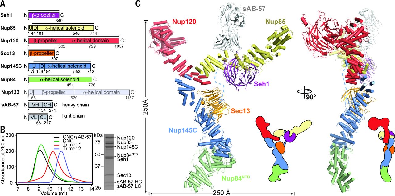
The nuclear pore complex (NPC) constitutes the sole gateway for bidirectional nucleocytoplasmic transport. Despite half a century of structural characterization, the architecture of the NPC remains unknown. In this research report, the authors present the crystal structure of a reconstituted ~400-kilodalton coat nucleoporin complex (CNC) from Saccharomyces cerevisiae at a 7.4 angstrom resolution. The crystal structure revealed a curved Y-shaped architecture and the molecular details of the coat nucleoporin interactions forming the central “triskelion” of the Y. A structural comparison of the yeast CNC with an electron microscopy reconstruction of its human counterpart suggested the evolutionary conservation of the elucidated architecture. Moreover, 32 copies of the CNC crystal structure docked readily into a cryoelectron tomographic reconstruction of the fully assembled human NPC, thereby accounting for ~16 megadalton of its mass.

Multilevel Summation Method for Electrostatic Force Evaluation

Authors: David J. Hardy, Zhe Wu, James C. Phillips, John E. Stone, Robert D. Skeel, and Klaus Schulten.

J Chem Theory Comput. 2015 Feb 10;11(2):766-779. PMCID: PMC4325600. (Consortium core: Computational Modeling Core)

Significant long-range electrostatic interactions arise in many biomolecular systems, such as negatively charged DNA and RNA, polar or charged membranes, ion channels, and electrostatic steering of protein−protein and enzyme−substrate association. Accordingly, electrostatic interactions need to be accurately represented in molecular modeling calculations. The computational cost increases in principle as N2, where N is the number of charged partiCl-es in the system.

The multilevel summation method (MSM) offers an efficient algorithm utilizing convolution for evaluating long-range forces arising in molecular dynamics simulations. Shifting the balance of computation and communication, MSM provides key advantages over the ubiquitous partiCl-e−mesh Ewald (PME) method, offering better scaling on parallel computers and permitting more modeling flexibility, with support for periodic systems as does PME but also for semiperiodic and nonperiodic systems. The version of MSM available in the simulation program NAMD is described, and its performance and accuracy are compared with the PME method. The accuracy feasible for MSM in practical applications reproduces PME results for water property calculations of density, diffusion constant, dielectric constant, surface tension, radial distribution function, and distance dependent Kirkwood factor, even though the numerical accuracy of PME is higher than that of MSM. Excellent agreement between MSM and PME is found also for interface potentials of air−water and membrane−water interfaces, where long-range Coulombic interactions are crucial. Applications demonstrate also the suitability of MSM for systems with semiperiodic and nonperiodic boundaries. For this purpose, simulations have been performed with periodic boundaries along directions parallel to a membrane surface but not along the surface normal, yielding membrane pore formation induced by an imbalance of charge across the membrane. Using a similar semiperiodic boundary condition, ion conduction through a graphene nanopore driven by an ion gradient has been simulated. Furthermore, proteins have been simulated inside a single spherical water droplet. Finally, parallel scalability results show the ability of MSM to outperform PME when scaling a system of modest size (less than 100 K atoms) to over a thousand processors, demonstrating the suitability of MSM for large-scale parallel simulation.

Ongoing is the development of improved interpolation for MSM to provide higher accuracy for a given polynomial degree p without increasing the computational cost. Future work inCl-udes also the calculation of dispersion forces without truncation with MSM-based NAMD; these forces, in particular, their long-range contribution, are considered to be important for membrane properties. With support in NAMD also for long-range dispersion forces, the present CHARMM-prescribed 12 Å cutoff/splitting distance can be used as a true control for MSM accuracy. High performance simulations will then be able to achieve practical accuracy with a reduced splitting distance, where a splitting distance of between 8 and 9 Å is expected to double the overall simulation performance.

Potential Application of Alchemical Free Energy Simulations to Discriminate GPCR Ligand Efficacy

Authors: Hui Sun Lee, Chaok Seok, and Wonpil Im.

J. Chem. Theory Comput. 2015 Feb 10;11:1255-1266. (Consortium core: Computational Modeling Core)

G protein-coupled receptors (GPCRs) constitute the largest protein superfamily in the human genome with almost 1,000  members. They play key functional roles as major contributors of information flow from the outside to the inside of the cell, making them one of the most important protein families. As a result of their broad influence on human physiology and behavior, GPCRs are arguably the most promising targets for development of new and more effective therapeutic agents.

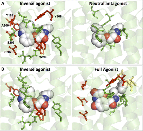
Based on the fact that GPCR-mediated signaling is modulated in a ligand-specific manner such as agonist, inverse agonist, and neutral antagonist (termed ligand efficacy), quantitative characterization of the ligand efficacy is essential for rational design of selective modulators for GPCR targets. As experimental approaches for this purpose are time-, cost-, and labor-intensive, computational tools that can systematically predict GPCR ligand efficacy can have a big impact on GPCR drug design. Here, the authors have performed free energy perturbation molecular dynamics simulations to calculate absolute binding free energy of an inverse agonist, a neutral antagonist, and an agonist to β2-adrenergic receptor (β2-AR) active and inactive states, respectively, in explicit lipid bilayers. Relatively short alchemical free energy calculations reveal that both the time series of the total binding free energy and decomposed energy contributions can be used as relevant physical properties to discriminate β2-AR ligand efficacy. This study illustrates a merit of the current approach over simple, fast docking calculations or highly expensive millisecond-time scale simulations.

It is the authors’ hope that their computational approach improves research and development efficiency in designing novel lead compounds targeting various GPCRs for the treatment of various human diseases.

Recent Consortium advances in the study of neurotransmitter transporters point the way to future research initiatives


Cartoon model of LeuT transport derived from EPR data, click to enlarge (Publication source)

Using the recently determined crystal structure of a prokaryotic leucine transporter (LeuT), the Transport Cycle in Neurotransmitter Uptake Project explores conformational changes and dynamic properties relevant to function in Neurotransmitter transporters translocation cycle using a combination of computational, functional, and spectroscopic approaches.

The conceptual design, scope and integration of this Project exemplifies the consortium approach to discovery of mechanistic principles of secondary active transport, including the conformational dynamics that govern alternating access in transporters and the allosteric interplay of substrate sites that transduce the energy stored in the electrochemical Na+ gradient into transport work. One of the most recent collaborative publications from members of this Project (lab of Harel Weinstein) and the Consortium at large (labs of Consortium member Olga Boudker and associate member Scott Blanchard) describes such conformational dynamics for a complex transporter system, illustrating the power of the combined experimental / computational approaches we have implemented for the study of these membrane proteins in the possible next phase of the grant:

Akyuz N, Georgieva ER, Zhou Z, Stolzenberg S, Cuendet MA, Khelashvili G, Altman RB, Terry DS, Freed JH, Weinstein H, Boudker O, Blanchard SC.
Transport domain unlocking sets the uptake rate of an aspartate transporter
Nature. 2015 Feb 5;518(7537):68-73. PMID: 25652997. PMCID: PMC4351760.


Crystal structure of the H276,395-GltPh.

Publication abstract: Glutamate transporters terminate neurotransmission by clearing synaptically released glutamate from the extracellular space, allowing repeated rounds of signalling and preventing glutamate-mediated excitotoxicity. Crystallographic studies of a glutamate transporter homologue from the archaeon Pyrococcus horikoshii, GltPh, showed that distinct transport domains translocate substrates into the cytoplasm by moving across the membrane within a central trimerization scaffold. Here we report direct observations of these ‘elevator-like’ transport domain motions in the context of reconstituted proteoliposomes and physiological ion gradients using single-molecule fluorescence resonance energy transfer (smFRET) imaging. We show that GltPh bearing two mutations introduced to impart characteristics of the human transporter exhibits markedly increased transport domain dynamics, which parallels an increased rate of substrate transport, thereby establishing a direct temporal relationship between transport domain motion and substrate uptake. Crystallographic and computational investigations corroborated these findings by revealing that the ‘humanizing’ mutations favour structurally ‘unlocked’ intermediate states in the transport cycle exhibiting increased solvent occupancy at the interface between the transport domain and the trimeric scaffold.

In a second publication from this Project, Matthias Quick (who is working with Project PI Jonathan Javitch), and Computational Modeling Core participant Lei Shi (from Harel Weinstein’s group), published findings that reveal the existence of two substrate sites in vSGLT, a member of the solute-sodium symporter (SSS) family of secondary transporters, as well as in PutP, another member of the SSS family. This publication follows a number of Consortium-sponsored interactions between Quick and Shi as well as Javitch and Weinstein. An earlier 2013 research initiative examined the aggregation dynamics and detergent binding to the second substrate site binding site of LeuT, and in the same year, their team published a key paper on the chloride binding site of neurotransmitter sodium transporters. The very first paper published in the name of the Project, was also the product of collaboration between this team of scientists, and showed how experimental conditions can block the second high-affinity site of LeuT. The important discovery of the present publication suggests that our findings in LeuT are generalizable to a different family of secondary transporters. These findings are the basis for forthcoming scientific collaborations between Quick and Shi, further demonstrating the broadening of the impact of the consortium and the synergy that the “glue” of the consortium has engendered.

Li Z, Lee AS, Bracher S, Jung H, Paz A, Kumar JP, Abramson J, Quick M, Shi L.
Identification of a second substrate-binding site in solute-sodium symporters
J Biol Chem., 2015 Jan 2;290(1):127-41. PMID: 25398883. PMCID: PMC4281715.


Crystallographically identified substrate-binding site of vSGLT is located more extracellularly than that of LeuT.

Publication abstract: The structure of the sodium/galactose transporter (vSGLT), a solute-sodium symporter (SSS) from Vibrio parahaemolyticus, shares a common structural fold with LeuT of the neurotransmitter-sodium symporter family. Structural alignments between LeuT and vSGLT reveal that the crystallographically identified galactose-binding site in vSGLT is located in a more extracellular location relative to the central substrate-binding site (S1) in LeuT. Our computational analyses suggest the existence of an additional galactose-binding site in vSGLT that aligns to the S1 site of LeuT. Radiolabeled galactose saturation binding experiments indicate that, like LeuT, vSGLT can simultaneously bind two substrate molecules under equilibrium conditions. Mutating key residues in the individual substrate-binding sites reduced the molar substrate-to-protein binding stoichiometry to ∼1. In addition, the related and more experimentally tractable SSS member PutP (the Na+/proline transporter) also exhibits a binding stoichiometry of 2. Targeting residues in the proposed sites with mutations results in the reduction of the binding stoichiometry and is accompanied by severely impaired translocation of proline. Our data suggest that substrate transport by SSS members requires both substrate-binding sites, thereby implying that SSSs and neurotransmitter-sodium symporters share common mechanistic elements in substrate transport.

CHARMM-GUI Membrane Builder publication by Im et al. featured as cover image for Journal of Computational Chemistry


Dr. Wonpil Im, University of Kansas

One of the ongoing projects in the laboratory of Wonpil Im (University of Kansas and MPSDC team member) has been to contribute to the development of CHARMM-GUI and CHARMM-GUI modules, such as the Ligand Binder and the Membrane Builder. These tools help users generate a series of CHARMM inputs towards a specific purpose, like calculating a protein/ligand binding free energy or building a protein/membrane complex for molecular dynamics simulations. The Consortium’s Computational Modeling Core has provided partial support for the completion of such modules.

Im and his team recently published a paper about the Membrane Builder module in Journal of Computational Chemistry, titled “CHARMM-GUI Membrane Builder toward realistic biological membrane simulations” (reference below). This publication was featured as the cover image for the issue, which can be viewed below. J Comput Chem attached the following note about the article’s relevance:

On page 1997 (DOI: 10.1002/jcc.23712), Emilia L. Wu, Xi Cheng, Sunhwan Jo, Huan Rui, Kevin C. Song, Eder M. Dávila‐Contreras, Yifei Qi, Jumin Lee, Viviana Monje‐Galvan, Richard M. Venable, Jeffery B. Klauda, and Wonpil Im report on the CHARMM‐GUI Membrane Builder, a web‐based user interface designed to interactively build all‐atom protein/membrane or membrane‐only systems for molecular dynamics simulation through an automated optimized process. The cover image illustrates the building process of a protein/membrane system by reading the protein structure, generating pore water/bilayer/bulk water/ions, and combining all the components.


(Click to enlarge)

Reference:

Wu EL, Cheng X, Jo S, Rui H, Song KC, Dávila-Contreras EM, Qi Y, Lee J, Monje-Galvan V, Venable RM, Klauda JB, Im W. CHARMM-GUI Membrane Builder toward realistic biological membrane simulations. J Comput Chem. 2014 Oct 15;35(27):1997-2004. PMID: 25130509. PMCID: PMC4165794 [Available on 2015/10/15].

Klaus Schulten’s keynote lecture movie “Photosynthetic Membrane of Purple Bacteria – A Clockwork of Proteins and Processes” made available with audio


Dr. Klaus Schulten

In the coming days, we will be releasing media footage from the exciting Frontiers in Membrane Protein Structural Dynamics 2014 meeting held at the Hilton Hotel in Chicago from May 7th-9th.

As a first piece of footage, we can think of no better than keynote speaker Klaus Schulten (UIUC)’s fascinating atom-by-atom movie titled “Photosynthetic Membrane of Purple Bacteria – A Clockwork of Proteins and Processes“, now made available with Schulten’s narrative of the movie during the keynote lecture.

We initially posted this as a silent movie to accompany our interview with professor Schulten in which he addresses his scientific research interests both past and present, his perspective on some of the key challenges for the field of membrane protein biophysics in the coming 5-10 years, his keynote lecture, and the Membrane Protein Structural Dynamics Consortium.

Now you can watch the movie together with Schulten’s own narration:

Collaborative LeuT study provides new view of neurotransmitter transporters

Model of LeuT alternating access inferred from the crystal structures.Model of LeuT alternating access inferred from the crystal structures.

This week, the Transport Cycle in Neurotransmitter Uptake Systems bridging project of the Membrane Protein Structural Dynamics Consortium (MPSDC) published an important article in Nature Structural & Molecular Biology on the bacterial leucine transporter (LeuT), a transporter which is structurally and functionally similar to neurotransmitter transporter proteins that direct neurotransmitters from synapse and terminal nerve signaling. The publication, titled “Conformational dynamics of ligand-dependent alternating access in LeuT,” was spearheaded by Vanderbilt graduate student Kelli Kazmier and Professor of Molecular Physiciology & Biophysics Hassane Mchaourab, and also featured collaboration by Consortium colleagues Jonathan Javitch, Harel Weinstein, and Benoît Roux.

The Transport Cycle in Neurotransmitter Uptake Systems project explores the conformational changes and dynamic properties relevant to function in Neurotransmitter transporters translocation cycle using a combination of computational, functional, and spectroscopic approaches. Using the recently determined crystal structure of a prokaryotic leucine transporter (LeuT), the scientists collaborating in this project are modeling the transport mechanisms of these proteins.

In this study, Mchaourab and colleagues used spectroscopic tools to make dynamic measurements in LeuT, in order to elucidate sodium- and leucine-dependent conformational This work highlights the importance of assessing the mechanistic identity of crystal structures, demonstrates the importance of dynamics in understanding function and realizes the vision of the consortium in integrating teams of scientists towards defining mechanistic principles of membrane proteins.changes in the transporter. The results identify the structural motifs that underlie the shift of LeuT between its various states – outward-facing, inward-facing and occluded. The conformational changes reported present a dynamic picture of the alternating-access mechanism of LeuT and NSSs that is different from the inferences reached from currently available structural models.

The publication marks a significant advance for the project’s research objectives, and is demonstrative of the cutting-edge collaborations between experimentalists and computationalists within the Consortium. According to Mchaourab, this work “highlights the importance of assessing the mechanistic identity of crystal structures, demonstrates the importance of dynamics in understanding function and realizes the vision of the consortium in integrating teams of scientists towards defining mechanistic principles of membrane proteins.”

The publication was also featured in Research News @ Vanderbilt. Click to read »

Interview with keynote speaker Klaus Schulten

Dr. Klaus Schulten Dr. Klaus Schulten

Three weeks in advance of Frontiers in Membrane Protein Structural Dynamics 2014, Professor Klaus Schulten (Director of the Computational and Theoretical Biophysics Group at UIUC) phoned in with us from his native Germany to talk about his research interests, the state of the field, the Membrane Protein Structural Dynamics Consortium (MPSDC), and the keynote lecture that he will be giving at the conference.

The full movie that will be shown in Klaus Schulten’s lecture can be viewed here.

Interviewer:
This is Rudo Kemper conducting an interview with Professor Klaus Schulten from the University of Illinois at Urbana-Champaign for the Membrane Protein Structural Dynamics Consortium.

Professor Schulten, please describe some of your scientific research interests, both past and present.

Klaus:
I’m a theoretical biophysicist and I try to approach living cells as a physicist to explore in how far cells exploit physical properties and physical laws and thereby to understand them better. My long goal is to achieve this by using theory as a kind of microscope and zoom through computer simulation into the processes in living cells; the views achieved this way are unobtainable for experiment, but in many ways can be verified by experiment. In other words, I wanted to build the world’s best microscope to view living cells and I sort of achieved it. Like any microscope, you can improve the computational microscope and reduce imaging artifacts, but by and large we can today image living cells through computer simulation with very high resolving power.

Photosynthetic chromatophore organelle (click for movie)

Interviewer:
In your view, what are some of the key challenges for the field of membrane protein biophysics in the coming 5-10 years?

Klaus:
One shouldn’t limit the question just to membrane biophysics because the membrane is only one part of the living cell. The cell has many components, the membrane is just one, and one has to see the cell as a whole rather than just focusing on one part. looking at small parts of the cell has been the dominant research focus for the last 50 years, looking basically at one protein at a time. In fact, biophysics had been extremely focused inside the cell looking at a volume that measured about 100 Angstroms x 100 Angstroms x 100 Angstroms at best, containing single proteins or protein complexes. But this cell volume is not where a biological cell really becomes alive.

The smallest living entity in Nature is a cell. A cell is 1 micrometer to 10 micrometers long in every direction, and so it is much bigger than the volume taken by single proteins or protein complexes. But somewhere between the small volume of a single protein and the large volume of the whole cell, life comes about. And this happens is what we want to understand, and we want to see what is it that makes life possible, that a cell can reproduce, can repair itself, organize itself, defend itself, live optimally in its environment. The point is actually rather simple: when you increasing the size of the volume of the cell that you visualize and thereby investigate, you may see at some volume size a very important phenomenon, namely how the many single molecules begin to cooperate in the cell and bring about the state that we call living. To appreciate this one needs to note that a living cell, for example a human cell, contains as many proteins as the United States has citizens. And so as have in our country many forms of organization, starting with the individual, then the family, then the neighborhood, the village, the city, the state and the country; there are also shops, companies, factories an army and more. And so likewise "social" interactions are also critical for the living cell and, in fact, it is the association and cooperation of molecules, for example proteins, what makes a cell alive. And so if you want to study how the living state develops, you have to study a volume of the cell that is much bigger than just what was studied before. Before, we studied phenomena in 100 Angstroms x 100 Angstroms x 100 Angstroms volume that contain up to about one million atoms. In the new era we seek to study volumes that contain up to a billion atoms. To visualize this large volume computationally we need to describe the detailed motion of as many atoms. That is a great challenge, but worth pursuing as we can learn then how biomolecules associate and cooperate in the cell, organize themselves, and thereby bring about life as we know it.

Interviewer:
What are you planning to talk about in your keynote lecture during the Frontiers in Membrane Proteins Structural Dynamics Conference in May?

LH1 – reaction center complex (click for movie)[caption align="alignright" width="300"] 
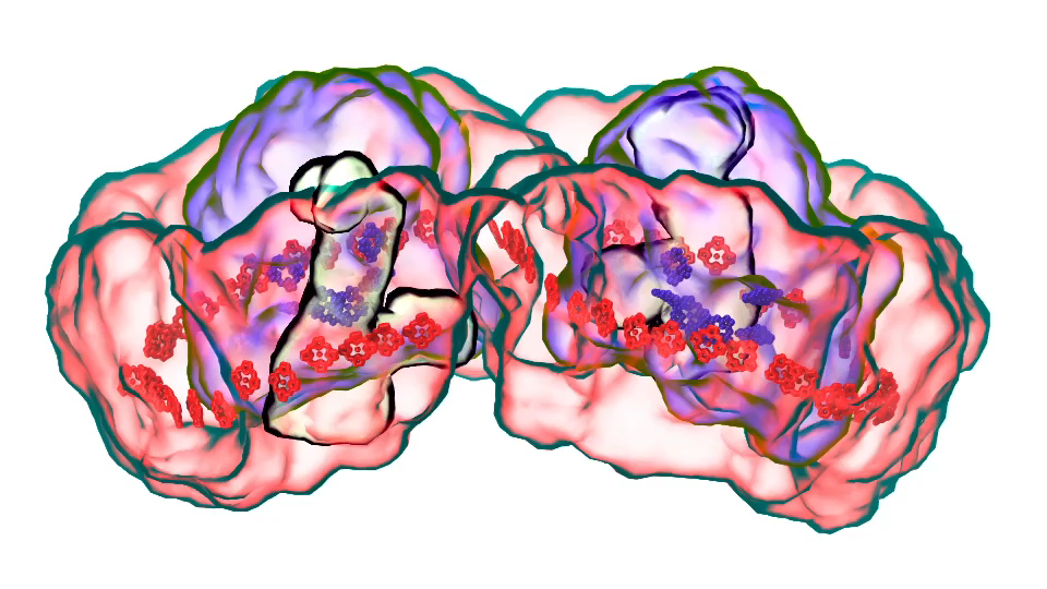
LH2 complex (click for movie)[/caption]

Klaus:
In Chicago I will talk about exactly one of those cases that I just mentioned. My talk is about a membrane system, called a chromatophore, that contains over a 100 million atoms a volume 700 Angstroms x 700 Angstroms x 700 Angstroms size. The chromatophore is a spherical membrane and will describe the processes that organize themselves in the chromatophore. The chromatophore is also called a cellular organelle and is found in huge numbers, like several hundreds, in certain photosynthetic bacteria, called purple bacteria. The chromatophore is a very critical organelle for the bacterium, because it gives the bacterium the energy it needs to sustain its life. It enables the bacterium to absorb sunlight and to turn it into chemical fuel that drives many processes in the cell. The key point of my lecture is that I will present the chromatophore as a whole. In the past we had been looking only at bits and pieces, at a single protein here, a light absorption process there. Now I show in my lecture that it is possible today to study the chromatophore, made of hundreds of proteins that permit many processes to run their course, as an entire system with all parts and processes included.

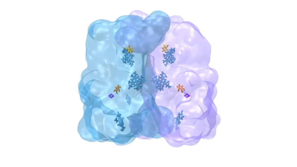
ATP synthase (click for movie)[caption align="alignright" width="300"] 
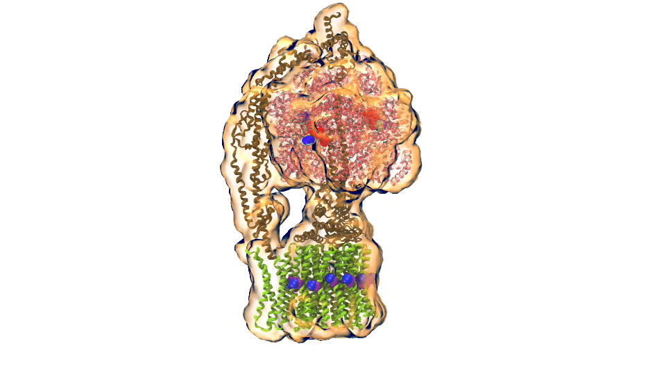
bc1 complex (click for movie)[/caption]

I had to actually work for 37 years of my life to get where I am and what I present in my lecture, and I’m not quite at the end even after so many years, but came far to give my lecture. The lecture will describe how all processes interlock with each other very much like the wheels making the horary in a Swiss watch. The processes (wheels) are all functioning together and work very precisely, just like a watch, despite thermal noise and other natural disorder. The very large biomolecular association that is the chromatophore has its architecture and processes perfectly attuned to permit purple bacteria that live mostly in very dim light, to nevertheless namely to make a living from the very few photons that exist in their habitat.

The chromatophore is composed of about 200 proteins and carries out about 30 processes. My lecture will start and end with an atom-by-atom movie, that is actually quite beautiful just like a movie in the theater, where one see all these processes taking place from the beginning when a photon is absorbed, all the way to the end when one sees the cellular fuel, molecules of ATP, being synthesized. One sees how the processes in the chromaotphore drive each other like a clockwork and are arranged beautifully together.

Interviewer:
In your view, how does the Glue Grant funded Membrane Protein Structural Dynamics Consortium contribute to the field?

Klaus:
The Consortium is contributing in two main respects to the field of membrane biology. On one side, it provides tools and laboratory capacities for membrane research. Membrane research is really very important, a key part of the cell is actually the membrane, many processes take place there, but it is a part of the cell that is much harder to study than the main part of the cell, called the cytosol. The Consortium develops tools that many other scientists can use, and it also provides collaborative capacities, so that researchers outside the Consortium can use the tools, coming to visit researchers who are members of the consortium, to collaborate with them and use their laboratory facilities.

This is one respect. The other respect is doing the most advanced research in membrane biology. For this purpose the consortium is constantly improving research tools so that the Consortium remains at the forefront of the field. Basically the mission of the Consortium is simple: do great science and at the same time develop new tools and make those tools available to everybody. This is a very, very important mission. The Consortium has a particularly strong computational side and, in keeping with its mission develops software that is used by many researchers in the United States and the world. The computational Consortium members have many researchers coming to visit for the investigation of membrane processes.

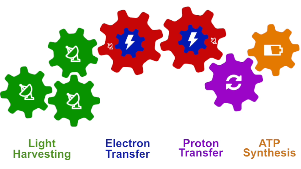
Photosynthesis – just like clockwork (click for movie)

Interviewer:
Finally, how have you and your team collaborated with some of the ongoing MPSDC projects?

Klaus:
My laboratory style is that basically all science projects are done together with experimentalists. As a result I benefit tremendously from the excellent experimental colleagues of the consortium. In the Consortium I have also really outstanding computational colleagues with whom I can develop new tools.

To be specific, we have one particular project where we study what is called the voltage sensing domain1. It is a very critical part of the so-called ion channels that exist, for example, in nerve cells, that have to adapt themselves to the spontaneous voltage of the nerve cell membrane. In this particular case, the experimentalists initially didn’t have the resolution power to get from their experimental observation the full atomic picture of the voltage sensing domain. But with a tool that my group developed for the purpose and tried out for the first time in this collaboration, we could actually develop a computationally resolve a key part of the atomic level structure. Later the experimentalist could actually solve the structure themselves, and we had a perfect agreement with the computational results so that we published together. We could also, as modelers, begin to explore the function of the voltage sensing domain. In any case we had a very close collaboration between computational and experimental membrane biologists.

1. Schulten has recently collaborated with the laboratories of Consortium colleagues Eduardo Perozo, Benoit Roux, and Anthony Kossiakoff to publish a paper titled Structural mechanism of voltage-dependent gating in an isolated voltage-sensing domain – an F1000Prime recommended article published in Nature Structural & Molecular Biology in March 2014

Klaus Schulten is Professor of Biophysics and Director of the Computational and Theoretical Biophysics Group at University of Illinois at Urbana-Champaign. Schulten is a leader in the field of computational biophysics, having devoted over 40 years to establishing the physical mechanisms underlying the processes and organization of living systems, from the atomic scale up to the level of the entire organism. As of 2014, his work in biological physics has yielded over 625 publications, which have been cited over 67,000 times. Schulten is also the co-director of the NSF-funded Center for the Physics of Living Cells and his work has been honored with numerous awards, including the Distinguished Service Award of the Biophysical Society in 2013, and the IEEE Computer Society Sidney Fernbach Award in 2012. Schulten also received the prestigious Humboldt Award of the German Humboldt Foundation in 2004.

Interview by Rudo Kemper, Web Content Administrator, Information and Dissemination Core

ABC Transporter paper by Tajkhorshid laboratory recommended on F1000Prime

One of our long-term projects on Structural Dynamics of ABC Transporters integrates computational, biochemical and spectroscopic approaches to understand the structural dynamics of the ATP binding cassette (ABC) transporters, which are associated with a number of human pathologies and play critical roles in the removal of cytotoxic agents.

Among the MPSDC participants in this project is Dr. Emad Tajkhorshid, whose contribution is applying molecular dynamics (MD) simulations integrating experimental constraints to develop structural models for key conformational states and characterize their inter-conversion during the transport cycle.

Tajkhorshid, together with postdoctoral researcher Mahmoud Moradi, recently published a paper on conformational transitions of ATP exporters which was recommended on the F1000Prime or “Faculty of 1000″ website. F1000 is a team of 5,000 Faculty Members – senior scientists and leading experts in all areas of biology and medicine — plus their associates who provide recommendations of important scientific articles, rating them and providing short explanations for their selections.

The publication by Moradi and Tajkhorshid, titled “Mechanistic picture for conformational transition of a membrane transporter at atomic resolution“, was published on November 19, 2013 in PNAS vol. 110, no. 47. The paper describes a nonequilibrium approach which they developed to characterize the conformational transition of MsbA, a member of the ATP-binding cassette exporter family, which is involved in transport of diverse substrates across the membrane. The F1000Prime recommendation, written by Qian Cui, tagged the publication as Good for Teaching, as having an Interesting Hypothesis, and for Technical Advance. Read the F1000 recommendation »

Dancing Proteins: Cell Membrane Transporter Motion May Revolutionize Drug Therapies (video)

The Beckman Institute at the University of Illinois at Urbana-Champaign produced the following video. In the video, Dr. Tajkhorshid describes how his laboratory has successfully simulated the molecular dance moves that a multidrug resistance membrane transporter undertakes as it pumps compounds out of a cell. This is the first time researchers have been able to simulate the motion of a complex membrane transporter in its native environment in full atomic detail and gives drug developers vital new targets to help combat drug-resistant cancers and other diseases.

Other plaudits

The popular NIH Biomedical Beat blog, which covers research news from NIGMS, featured the video on their website. As per the blog page, “In this video, Emad Tajkhorshid of the University of Illinois at Urbana-Champaign explains the molecular dance of ABC transporters, a family of molecular machines that utilize ATP to move substances across the cell membrane. Tajkhorshid and his team recently used computational methods to map the movements between two known structural models of MsbA, a bacterial version of a transporter in human cells that helps to export anti-cancer drugs. They then described the individual steps of the molecular motions during the transport cycle. Understanding the process at such a detailed level could suggest new targets for treating a range of diseases, including some drug-resistant cancers that often make more transporter proteins to kick out medications meant to kill them.”

Additionally, Tajkhorshid and Moradi were also featured in the University of Illinois News Bureau, in an article titled “Difficult dance steps: Team learns how membrane transporter moves.” The article helpfully describes the nature of the research and points to its innovation.


Photo taken from the University of Illinois News Bureau website. Photo by L. Brian Stauffer.

According to the article, “the new findings, reported in the Proceedings of the National Academy of Sciences, will help scientists figure out how other transporters work. The work also offers new insights into multi-drug-resistant (MDR) cancers, some of which use these transporters to export cancer-killing drugs.” Previously, it has been difficult to research large, membrane-bound proteins like MsbA because they are not easy to crystallize, and each crystal structure reflects only one of the many conformations of these shape-shifting proteins. This study marks “the first time that we are characterizing a very complex structural transition at atomic-level resolution for a large protein,” Dr. Tajkhorshid is quoted as saying.

Emad Tajkhorshid is Professor of Biochemistry, Biophysics, and Pharmacology and an affiliate of the Beckman Institute for Advanced Science and Technology at the University of Illinois at Urbana-Champaign.

Categories
Recent Posts
Archives
Recent Comments
Recent Photos