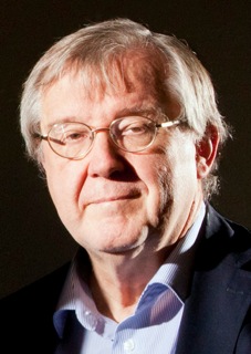
Behind the Scenes

Klaus Schulten’s keynote lecture movie “Photosynthetic Membrane of Purple Bacteria – A Clockwork of Proteins and Processes” made available with audio


Dr. Klaus Schulten

In the coming days, we will be releasing media footage from the exciting Frontiers in Membrane Protein Structural Dynamics 2014 meeting held at the Hilton Hotel in Chicago from May 7th-9th.

As a first piece of footage, we can think of no better than keynote speaker Klaus Schulten (UIUC)’s fascinating atom-by-atom movie titled “Photosynthetic Membrane of Purple Bacteria – A Clockwork of Proteins and Processes“, now made available with Schulten’s narrative of the movie during the keynote lecture.

We initially posted this as a silent movie to accompany our interview with professor Schulten in which he addresses his scientific research interests both past and present, his perspective on some of the key challenges for the field of membrane protein biophysics in the coming 5-10 years, his keynote lecture, and the Membrane Protein Structural Dynamics Consortium.

Now you can watch the movie together with Schulten’s own narration:

Interview with keynote speaker Klaus Schulten

Dr. Klaus Schulten Dr. Klaus Schulten

Three weeks in advance of Frontiers in Membrane Protein Structural Dynamics 2014, Professor Klaus Schulten (Director of the Computational and Theoretical Biophysics Group at UIUC) phoned in with us from his native Germany to talk about his research interests, the state of the field, the Membrane Protein Structural Dynamics Consortium (MPSDC), and the keynote lecture that he will be giving at the conference.

The full movie that will be shown in Klaus Schulten’s lecture can be viewed here.

Interviewer:
This is Rudo Kemper conducting an interview with Professor Klaus Schulten from the University of Illinois at Urbana-Champaign for the Membrane Protein Structural Dynamics Consortium.

Professor Schulten, please describe some of your scientific research interests, both past and present.

Klaus:
I’m a theoretical biophysicist and I try to approach living cells as a physicist to explore in how far cells exploit physical properties and physical laws and thereby to understand them better. My long goal is to achieve this by using theory as a kind of microscope and zoom through computer simulation into the processes in living cells; the views achieved this way are unobtainable for experiment, but in many ways can be verified by experiment. In other words, I wanted to build the world’s best microscope to view living cells and I sort of achieved it. Like any microscope, you can improve the computational microscope and reduce imaging artifacts, but by and large we can today image living cells through computer simulation with very high resolving power.

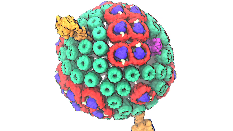
Photosynthetic chromatophore organelle (click for movie)

Interviewer:
In your view, what are some of the key challenges for the field of membrane protein biophysics in the coming 5-10 years?

Klaus:
One shouldn’t limit the question just to membrane biophysics because the membrane is only one part of the living cell. The cell has many components, the membrane is just one, and one has to see the cell as a whole rather than just focusing on one part. looking at small parts of the cell has been the dominant research focus for the last 50 years, looking basically at one protein at a time. In fact, biophysics had been extremely focused inside the cell looking at a volume that measured about 100 Angstroms x 100 Angstroms x 100 Angstroms at best, containing single proteins or protein complexes. But this cell volume is not where a biological cell really becomes alive.

The smallest living entity in Nature is a cell. A cell is 1 micrometer to 10 micrometers long in every direction, and so it is much bigger than the volume taken by single proteins or protein complexes. But somewhere between the small volume of a single protein and the large volume of the whole cell, life comes about. And this happens is what we want to understand, and we want to see what is it that makes life possible, that a cell can reproduce, can repair itself, organize itself, defend itself, live optimally in its environment. The point is actually rather simple: when you increasing the size of the volume of the cell that you visualize and thereby investigate, you may see at some volume size a very important phenomenon, namely how the many single molecules begin to cooperate in the cell and bring about the state that we call living. To appreciate this one needs to note that a living cell, for example a human cell, contains as many proteins as the United States has citizens. And so as have in our country many forms of organization, starting with the individual, then the family, then the neighborhood, the village, the city, the state and the country; there are also shops, companies, factories an army and more. And so likewise "social" interactions are also critical for the living cell and, in fact, it is the association and cooperation of molecules, for example proteins, what makes a cell alive. And so if you want to study how the living state develops, you have to study a volume of the cell that is much bigger than just what was studied before. Before, we studied phenomena in 100 Angstroms x 100 Angstroms x 100 Angstroms volume that contain up to about one million atoms. In the new era we seek to study volumes that contain up to a billion atoms. To visualize this large volume computationally we need to describe the detailed motion of as many atoms. That is a great challenge, but worth pursuing as we can learn then how biomolecules associate and cooperate in the cell, organize themselves, and thereby bring about life as we know it.

Interviewer:
What are you planning to talk about in your keynote lecture during the Frontiers in Membrane Proteins Structural Dynamics Conference in May?

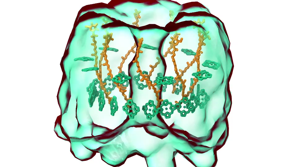
LH1 – reaction center complex (click for movie)[caption align="alignright" width="300"] 
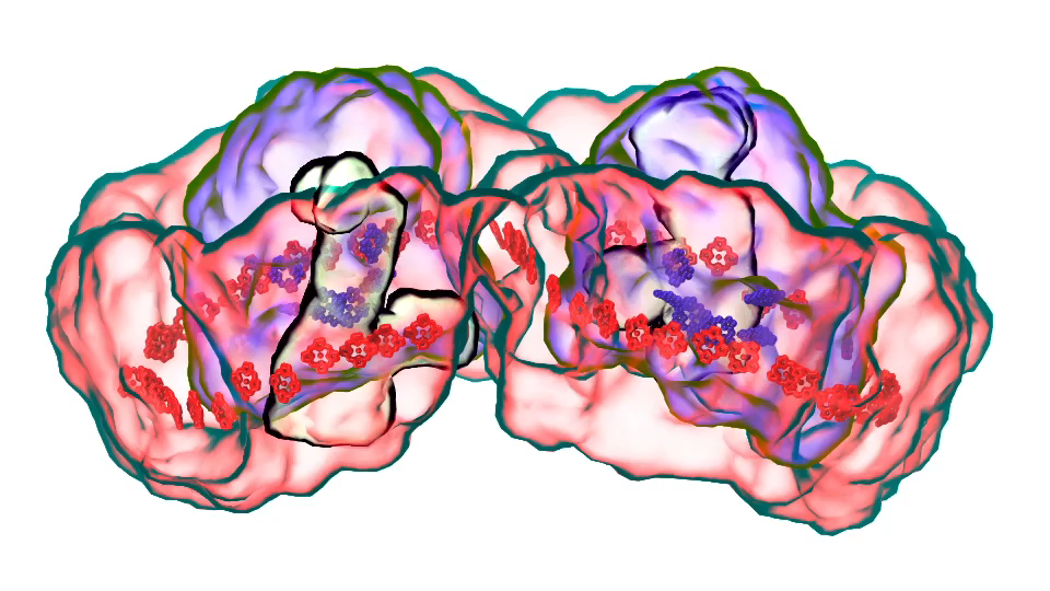
LH2 complex (click for movie)[/caption]

Klaus:
In Chicago I will talk about exactly one of those cases that I just mentioned. My talk is about a membrane system, called a chromatophore, that contains over a 100 million atoms a volume 700 Angstroms x 700 Angstroms x 700 Angstroms size. The chromatophore is a spherical membrane and will describe the processes that organize themselves in the chromatophore. The chromatophore is also called a cellular organelle and is found in huge numbers, like several hundreds, in certain photosynthetic bacteria, called purple bacteria. The chromatophore is a very critical organelle for the bacterium, because it gives the bacterium the energy it needs to sustain its life. It enables the bacterium to absorb sunlight and to turn it into chemical fuel that drives many processes in the cell. The key point of my lecture is that I will present the chromatophore as a whole. In the past we had been looking only at bits and pieces, at a single protein here, a light absorption process there. Now I show in my lecture that it is possible today to study the chromatophore, made of hundreds of proteins that permit many processes to run their course, as an entire system with all parts and processes included.

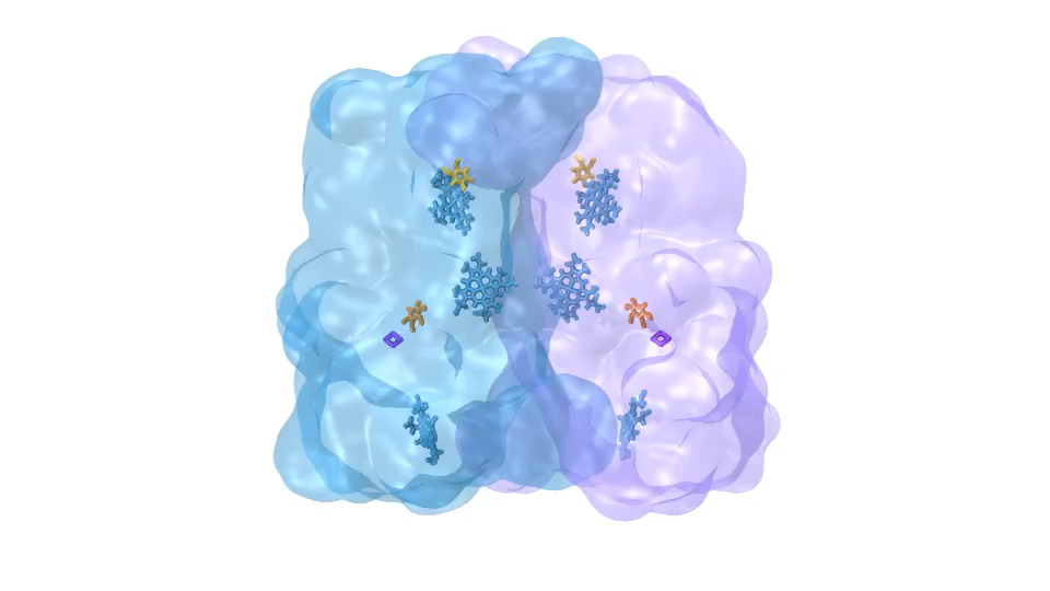
ATP synthase (click for movie)[caption align="alignright" width="300"] 
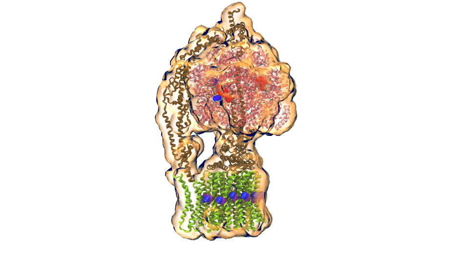
bc1 complex (click for movie)[/caption]

I had to actually work for 37 years of my life to get where I am and what I present in my lecture, and I’m not quite at the end even after so many years, but came far to give my lecture. The lecture will describe how all processes interlock with each other very much like the wheels making the horary in a Swiss watch. The processes (wheels) are all functioning together and work very precisely, just like a watch, despite thermal noise and other natural disorder. The very large biomolecular association that is the chromatophore has its architecture and processes perfectly attuned to permit purple bacteria that live mostly in very dim light, to nevertheless namely to make a living from the very few photons that exist in their habitat.

The chromatophore is composed of about 200 proteins and carries out about 30 processes. My lecture will start and end with an atom-by-atom movie, that is actually quite beautiful just like a movie in the theater, where one see all these processes taking place from the beginning when a photon is absorbed, all the way to the end when one sees the cellular fuel, molecules of ATP, being synthesized. One sees how the processes in the chromaotphore drive each other like a clockwork and are arranged beautifully together.

Interviewer:
In your view, how does the Glue Grant funded Membrane Protein Structural Dynamics Consortium contribute to the field?

Klaus:
The Consortium is contributing in two main respects to the field of membrane biology. On one side, it provides tools and laboratory capacities for membrane research. Membrane research is really very important, a key part of the cell is actually the membrane, many processes take place there, but it is a part of the cell that is much harder to study than the main part of the cell, called the cytosol. The Consortium develops tools that many other scientists can use, and it also provides collaborative capacities, so that researchers outside the Consortium can use the tools, coming to visit researchers who are members of the consortium, to collaborate with them and use their laboratory facilities.

This is one respect. The other respect is doing the most advanced research in membrane biology. For this purpose the consortium is constantly improving research tools so that the Consortium remains at the forefront of the field. Basically the mission of the Consortium is simple: do great science and at the same time develop new tools and make those tools available to everybody. This is a very, very important mission. The Consortium has a particularly strong computational side and, in keeping with its mission develops software that is used by many researchers in the United States and the world. The computational Consortium members have many researchers coming to visit for the investigation of membrane processes.

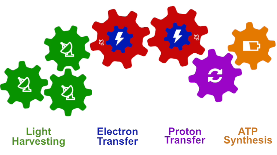
Photosynthesis – just like clockwork (click for movie)

Interviewer:
Finally, how have you and your team collaborated with some of the ongoing MPSDC projects?

Klaus:
My laboratory style is that basically all science projects are done together with experimentalists. As a result I benefit tremendously from the excellent experimental colleagues of the consortium. In the Consortium I have also really outstanding computational colleagues with whom I can develop new tools.

To be specific, we have one particular project where we study what is called the voltage sensing domain1. It is a very critical part of the so-called ion channels that exist, for example, in nerve cells, that have to adapt themselves to the spontaneous voltage of the nerve cell membrane. In this particular case, the experimentalists initially didn’t have the resolution power to get from their experimental observation the full atomic picture of the voltage sensing domain. But with a tool that my group developed for the purpose and tried out for the first time in this collaboration, we could actually develop a computationally resolve a key part of the atomic level structure. Later the experimentalist could actually solve the structure themselves, and we had a perfect agreement with the computational results so that we published together. We could also, as modelers, begin to explore the function of the voltage sensing domain. In any case we had a very close collaboration between computational and experimental membrane biologists.

1. Schulten has recently collaborated with the laboratories of Consortium colleagues Eduardo Perozo, Benoit Roux, and Anthony Kossiakoff to publish a paper titled Structural mechanism of voltage-dependent gating in an isolated voltage-sensing domain – an F1000Prime recommended article published in Nature Structural & Molecular Biology in March 2014

Klaus Schulten is Professor of Biophysics and Director of the Computational and Theoretical Biophysics Group at University of Illinois at Urbana-Champaign. Schulten is a leader in the field of computational biophysics, having devoted over 40 years to establishing the physical mechanisms underlying the processes and organization of living systems, from the atomic scale up to the level of the entire organism. As of 2014, his work in biological physics has yielded over 625 publications, which have been cited over 67,000 times. Schulten is also the co-director of the NSF-funded Center for the Physics of Living Cells and his work has been honored with numerous awards, including the Distinguished Service Award of the Biophysical Society in 2013, and the IEEE Computer Society Sidney Fernbach Award in 2012. Schulten also received the prestigious Humboldt Award of the German Humboldt Foundation in 2004.

Interview by Rudo Kemper, Web Content Administrator, Information and Dissemination Core

Interview with Robert Nakamoto, MPSDC collaborator and Chair of the 2014 BPS Program Committee

MPSDC Team Member Robert Nakamoto, who leads the Consortium’s Protein Production/Expression Core, is this year’s Chair of the 2014 Biophysical Society Program Committee. Nakamoto was interviewed several times by Biophysical Society TV for the occasion of the 2014 Biophysical Society meetings. You can check out the interviews below:

Interview with Francisco Bezanilla, MPSDC collaborator and President of the Biophysical Society

Biophysical Society president and MPSDC Team Member Francisco Bezanilla was interviewed today by Biophysical Society TV. You can check out the interview below:

Video feature: Behind the scenes in Emad Tajkhorshid’s laboratory

We are delighted to share with you the latest episode of our Behind the Scenes series. Within this series, we explore the inner workings of the Consortium with a camera in hand, dedicating to showing you what takes place within the laboratory setting and outside of it.

For our second feature, we present the following video feature of Emad Tajkhorshid, an active participant in the Consortium’s Computational Modeling Core and the Structural Dynamics of ABC Transporter and Conformational Dynamics in the CLC Channel/Transporter Family projects. We sat down with Emad and asked him about his work with computational modeling, how his laboratory collaborates with other entities within the Consortium, and how he views the role of the Consortium within the broader scientific community. Let us know what you think in the comments!

Consortium progress and scientific advances discussed at third annual meeting

2013 group photoClick to enlarge.

For the third year in a row, the Membrane Protein Structural Dynamics Consortium (MPSDC) hosted a number of events in its home base of Chicago during the month of May, key among these its third annual meeting. Unlike last year’s Frontiers in Membrane Protein Structural Dynamics conference, this year’s annual meeting was closed to the public, although there are plans to host a second open attendance conference in the near future.

Core workshops and minisymposium

As in previous years, the MPSDC’s Computational Modeling Core hosted a membrane protein modeling workshop, and a mini-symposium concerning the latest advances in computational approaches to the study of membrane proteins. As before, the modeling workshop provided attendants with an overview of the use of the modeling dynamics and visualization software NAMD and VMD, and also featured Dr. Wonpil Im’s CHARMM-GUI Ligand Binder module. This year’s mini-symposium covered a number of topics including force field and atomic models, structural modeling with low-resolution data, and transition pathways. The minisymposium hosted a “keynote” lecture of sorts on 2D-IR Spectroscopy, held by Josh Carr from the University of Wisconson-Madison. We are pleased to present you with a recording of Carr’s lecture, titled Connecting Experiment and Simulation by Modeling the Protein Amide I Band.

This year, the Consortium’s Membrane Protein Expression/Purification Core held its first workshop as well. This workshop featured several Consortium collaborators such as Edith Buchinger from Goethe University, Stephen Pless and Lilie Leisle from the University of Iowa, and Andrzej Rajca from the University of Nebraska. Topics discussed at this workshop included cellular and cell-free production of membrane proteins, reconstitution, incorporation of unnatural amino acids, single antigen binder technologies, and chemistry of protein modification and nitroxide spin labels. Both workshops and minisymposium were well attended and productive, and we will continue to host such satellite events in the future.

Annual meeting

The MPSDC’s annual meeting allows for Consortium PI’s to present on the latest advances in the respective cores and projects, and serves as a dedicated time and space for members to discuss their research and organically find ways to collaborate with colleagues. Moreover, it gives the executive arm of the MPSDC the opportunity to report on “the state of the Consortium.”

At this year’s annual meeting at the University of Chicago’s Gleacher Center, MPSDC director Eduardo Perozo highlighted the continual exchange of ideas within and dynamics of the Consortium itself. Focusing specifically on the activities of the past year’s accomplishments, Dr. Perozo discussed the rational and efficient consolidation of Core Facilities to their highest efficiency and optimal productivity in close collaboration with individual projects. For example, the Computational Modeling core is starting to provide a number of services and home-developed algorithms to the community at large, from quick force field parameterization of small molecules (and potential membrane protein ligands) to the conformational energetics biophysical probes, to important tools to the use and interpretation of long range distances and distance distributions from DEER experiments. Additionally, Dr. Perozo reported on the status of the Consortium’s unique bridging and pilot projects. Four of our bridging projects are currently in full swing, and one of the pilot projects has successfully transitioned to bridging project status. We have also incorporated two exciting new Pilot projects that bring new systems and new techniques to the Consortium.

Followed by Dr. Perozo’s framing discussion, the PI’s of each of the three cores and seven projects described the progress made and latest scientific findings in their respective teams, providing attendees with the opportunity to respond and provide helpful feedback. The cores are designed to act as “innovation incubators” and research support centers by providing service and expertise in these critical areas: Membrane protein expression, the establishment of chemical synthesis capabilities for probes and detergents, the generation of a variety of binders and other crystallization chaperones and other target binders and the development of common computational tools to interpret and integrate the wealth of experimental data. Each of these feed and interconnect with individual projects in a highly interactive way.

The Consortium’s projects are integrated as research efforts that tie together or enhance the contribution of the independent work and expertise of the participating investigator to the Consortium and expand the independent work in new directions. Ten talks in all, each of the PI’s of the MPSDC’s cores and projects presented on the very latest activities, which will soon be disseminated to the public in the form of publications and resources.

On the second day of the annual meeting, we featured presentations from a number of our associate members. Associate members are junior faculty members who are pursuing exciting and innovative research parallel to the Consortium’s mission, and have therefore been given access to the interactions and core resources of the Consortium in a budget-neutral way.

Several of our associate members have gone on to participate full-fledged in the Consortium’s activities, either by way of participating in existing projects (such as Wonpil Im with the Computational Modeling Core or Chris Ahern with the Membrane Protein Expression/Purification Core) or by starting new pilot projects. Both Olga Boudker and Ming Zhou started as associated members, and after presenting on their research at last year’s conference have gone on to spearhead new and promising pilot projects.

Along these lines, we invited several of our associate members to present talks, in order to foster further cross-pollination between our research and theirs. This year, Luis Cuello from Texas Tech University presented on inactiviation gating at the K+ channel selectivity filter, Katherine Henzler-Wildman from Washington University presented on the mechanism of multi drug efflux by EmrE, Jens Meiler from Vanderbilt University presented on membrane protein structure & dynamics from limited experimental data, and Robert Keenan from the University of Chicago gave a talk on tail-anchored membrane protein insertion at the ER. In addition to Andrzej Rajca who participated in the Membrane Protein Production core workshop and other associate members like Francis Valiyaveetil, all of them bring state of the art expertise in different areas such as synthetic chemistry and chemical biology, and are expected to actively participate in several of the Consortium’s activities.

In sum, we are thrilled to report that the Consortium is thriving, from both a strictly scientific stand point as well as in regards to our output to the community. We’d like to thank all who attended and partook in this year’s discussions, and look forward to seeing you next year!

Below are several photos of the annual meeting and satellite events. You can either browse through the photos here or visit the set on Flickr.

First Frontiers in Membrane Protein Structural Dynamics Conference was a success


Dorothee Kern, Brandeis University and External Advisory Committee Member

On May 3rd and 4th, the Membrane Protein Structural Dynamics Consortium (MPSDC) held its first Frontiers in Membrane Protein Structural Dynamics conference. The conference consisted of scientific sessions and poster presentations, and featured both Consortium members and external invitees. Attendance was open to the public took place within the context of our 3rd Annual MPSDC Meeting, where all members, NIH representatives and our External Advisory Committee participated. Prior to the conference, the MPSDC’s Computational Modeling Core hosted a NAMD/VMD workshop and a mini-symposium concerning the latest advances in membrane protein modeling.


Miguel Holmgren, NINDS and collaborator in Bridge 1: Conformational Transitions in P-class ATPases

Both the conference and CMC events were very well attended and enabled extensive conversations surrounding the topic of cutting edge advances and scientific methods in the field of membrane protein dynamics, as well as ways to resolve current roadblocks. The conference was able to hit a high note in bringing together the issues and ideas most relevant to the key goals of the consortium both in the present and in the future. Chris Ahern of the University of British Columbia and MPSDC Associate Member noted afterwards that it was probably one of the best meetings he’s been to, “primarily because of the quality of the science that’s being done, as well as the excitement and eagerness of people to cooperate.”


José Faraldo-Gómez, Max Planck Institute for Biophysics and MPSDC Associate Member

In the afternoon, two discussion panels were held, which themselves are in the spirit of much of the broader conversations that took place at the conference as a whole. The first panel, “Finding a common language: linking experiment and computation” was chaired by Hassane Mchaourab, and included Benoît Roux, Martin Zanni, Ivet Bahar, Dorothee Kern, and Emad Tajkhorshid as participants.


Chris Ahern, University of British Columbia and MPSDC Associate Member

The second panel was titled “Breaking the barriers of membrane protein expression and labeling” and was moderated by Robert Nakamoto. The panel included Chris Ahern, Jim Bowie, Volker Dötsch and Shohei Koide. Discussion focused on three topics: the optimal nitroxide spin probe for monitoring protein dynamics and DEER distance measurements, how to incorporate such a probe into the protein targets, and cell free synthesis of target proteins. The optimal nitroxide spin probe would be connected to the backbone by only a β carbon. Such a label can be introduced using chemical synthesis, but because most of our proteins are too large, methods for bio-incorporation of unnatural amino acid are preferred. Cell free biosynthesis systems such as those developed in the laboratory of Volker Dötsch, Goethe University, and Chris Ahern, University of British Columbia, may provide the best approaches. The Protein Core labs will explore methods for charging the non-sense tRNA by evolved tRNA synthetases or a ribozyme using technologies called the Flexizyme developed by Soga and co-workers at Tokyo University. The acylated TAG tRNA charged with the spin probe is simply added to the cell free synthesis mix. Another issue is the lability of nitoxides to reduction by ascorbic acid. We will test a variety of nitroxide spin probes, which have been reported to be relatively insensitive to reduction. Finally, we discussed specific placement of labels using synthetic binders. In particular, the synthetic 10FN3 binders, or monobodies (~93 aa, derived from a type III fibronectin domain), can mediate specific labeling of a protein and effectively attach a label or cargo to the protein target with high affinity and stability.

We’d like to thank all who attended and partook in the discussions. By all accounts, it is hard to think of a better outcome for the conference and accompanying events, and we look forward to hosting another meeting in two years.

Below is a gallery displaying photos of the conference. You can either scroll through the photos here or visit the set on Flickr. We’ve also made available several brief audio interviews with attendees of the conference, to be found in the margins of the body of this post.

Benoît Roux and the Computational Modeling Core

We are delighted to introduce the latest feature on the Membrane Protein Structural Dynamics Consortium (MPSDC) website: Behind the Scenes. Within this section, we will be exploring the inner workings of the Consortium with a camera in hand, dedicating to showing you what takes place within the laboratory setting and outside of it.

For our inaugural piece, we present the following video feature of Benoît Roux, the PI of the Consortium’s Computational Modeling Core (CMC). We sat down with Benoît and asked him about computational modeling, the CMC’s collaborations and function within the MPSDC, and where he sees the Consortium in five years. Let us know what you think in the comments!

Categories
Recent Posts
Archives
Recent Comments
Recent Photos