Molecular insights into the membrane-associated phosphatidylinositol 4-kinase IIα
By Qiangjun Zhou, Jiangmei Li, Hang Yu, Yujia Zhai, Zhen Gao, Yanxin Liu, Xiaoyun Pang, Lunfeng Zhang, Klaus Schulten, Fei Sun and Chang Chen.
Published in Nature Communications 5(3552) on March 28, 2014. PMID: 24675427. Link to publication page.
Core Facility: Computational Modeling
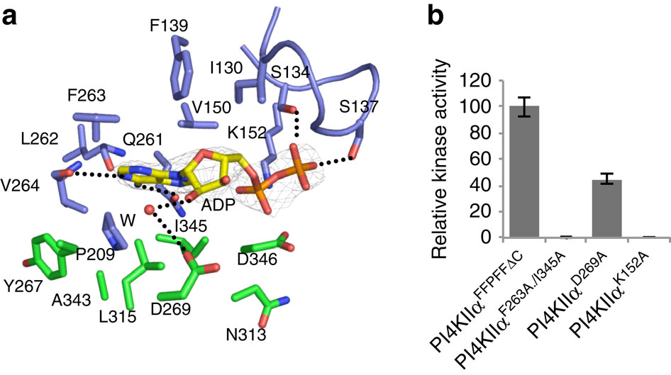
Figure 2. Nucleotide-binding pocket of PI4KIIα. (a) Interactions between ADP and PI4KIIα. Residues involved in ADP binding are labelled and shown as stick models with those from the N-lobe coloured in blue and those from the C-lobe coloured in green. A water molecule (W, red) is shown in sphere. The 2Fo-Fc electron density map is shown as a grey mesh and contoured at 1.8σ. Hydrogen bonds are shown as dashed lines. See also Table 2. (b) Kinase activities of PI4KIIα variants with mutations at the nucleotide-binding site. Kinase activity was measured in PI/Triton X-100 (0.2%) and normalized by using the activity of PI4KIIαFFPFFDC as 100%. The error bars represent the s.d. from three independent experiments.
Abstract
Phosphatidylinositol 4-kinase IIα (PI4KIIα), a membrane-associated PI kinase, plays a central role in cell signalling and trafficking. Its kinase activity critically depends on palmitoylation of its cysteine-rich motif (-CCPCC-) and is modulated by the membrane environment. Lack of atomic structure impairs our understanding of the mechanism regulating kinase activity. Here we present the crystal structure of human PI4KIIα in ADP-bound form. The structure identifies the nucleotide-binding pocket that differs notably from that found in PI3Ks. Two structural insertions, a palmitoylation insertion and an RK-rich insertion, endow PI4KIIα with the ‘integral’ membrane-binding feature. Molecular dynamics simulations, biochemical and mutagenesis studies reveal that the palmitoylation insertion, containing an amphipathic helix, contributes to the PI-binding pocket and anchors PI4KIIα to the membrane, suggesting that fluctuation of the palmitoylation insertion affects PI4KIIα’s activity. We conclude from our results that PI4KIIα’s activity is regulated indirectly through changes in the membrane environment.
 Primary structure of PI4KIIα. I1, I2 and I3 represent three insertions in PI4KIIα not found in PI3Ks. Palmitoylation and RK-rich insertions are referred as I1 and I2, respectively. The crystallized fragment PI4KIIαSSPSSΔC is indicated by a black arrow. (b) Purification and kinase activity assay of PI4KIIαSSPSSΔC. The kinase activity of PI4KIIαSSPSSΔC in PI/Triton X-100 (0.2%)(▴) or PI-containing liposome/Triton X-100 (0.2%)(▪) was measured by monitoring ADP production. The error bars represent the s.d. from three independent experiments. (c) Overall structure of the ADP-bound PI4KIIα catalytic domain. Untraced segments are depicted with dashed lines. The G-loop, catalytic loop and activation loop are coloured in pink, bright orange and magenta, respectively. The palmitoylation insertion (PAL insertion, I1) and RK-rich insertion (I2) are coloured in red and gold, respectively. ADP is shown in a sphere model and is coloured according to its atoms (carbon, yellow; oxygen, red; nitrogen, blue; phosphorus, orange). The colour scheme here is used in all figures unless otherwise mentioned. (d) Secondary structure topology of the PI4KIIα catalytic domain. All structural figures were prepared with PyMol (DeLano, 2002, http://www.pymol.org) or UCSF Chimera ( http://www.cgl.ucsf.edu.libproxy.lib.unc.edu/chimera/).")
 Sequential membrane elution assay to evaluate the membrane binding of PI4KIIα variants. (c) The membrane-binding surface of PI4KIIα deduced from the crystallographic packing analysis and mapped with its electrostatic potentials. Blue represents positive charges and red represents negative charges. ADP and the putative PI (see Fig. 4b) are shown as stick models. Residues potentially involved in membrane binding are indicated accordingly. (d) Kinase activities of the PI4KIIα variants. The kinase activity was measured in PI/Triton X-100 (0.2%) and monitored by ADP production. The error bars represent the s.d. from three independent experiments. (e) A representative model of PI4KIIα bound to membrane, obtained from MD simulations. The crystal structure is coloured in grey and the MD structural model is shown with the same scheme of Fig. 1c. The residues evaluated in (d) are indicated accordingly. The simulated membrane is depicted in transparent dots. The left panel is perpendicular to the membrane and the right one is parallel and viewed from the membrane side.")
 Putative PI-binding pocket of PI4KIIα based on molecular dynamics simulations. The cavity surrounded by the palmitoylation insertion and activation loop was computed by using HOLLOW39 and rendered in brown surface. (b) Identified important residues for contributing to the PI-binding pocket. These residues are labelled, coloured in yellow and mapped onto the surface of PI4KIIα. The structural model of PI4KIIα including ATP and PI substrates is derived from a snapshot of molecular dynamics simulation. (c) Kinase activities of PI4KIIα variants with mutations at the putative PI-binding pocket. The kinase activity was measured in PI/Triton X-100 (0.2%) and normalized by using the activity of PI4KIIαCCPCC as 100%. All error bars here represent the s.d. from three independent experiments. (d) The kinase activities of PI4KIIα variants with mutations at the amphipathic helix of the palmitoylation insertion. The kinase activity was measured in PI/0.1% Anzergent3-14 and normalized by using the activity of the construct PI4KIIαCCPCC as 100%. The error bars in (c) and (d) represent the s.d. from three independent experiments. (e,f) Conformational fluctuations of PI4KIIα without palmitoylation in (e) and with palmitoylation in (f). Proteins were first aligned to the conformations at t=300 ns in each trajectory using backbone atoms of proteins not including three insertions (I1, I2 and I3). The root mean square fluctuation (RMSF) was then calculated using C-alpha atoms of all residues (residue 107 to 453) over the last 700 ns. Blue, green and dark green curves in (e) represent the RMSF statistics from three independent simulations of PI4KIIα without palmitoylation. Orange, red and pink curves in (f) represent three independent simulation of palmitoylated PI4KIIα.")
