On the structural basis of modal gating behavior in K(+) channels
By Sudha Chakrapani, Julio F Cordero-Morales, Vishwanath Jogini, Albert C Pan, D Marien Cortes, Benoît Roux, and Eduardo Perozo
Published in Nature Structural & Molecular Biology 18(1): 67-74 on Jan, 2011.
PMID: 21186363. PMCID: PMC3059741. Link to Pubmed page.
Project: Dynamics of Ion Permeation and Conformational Coupling in – KcsA. Core Facility: Computational Modeling.
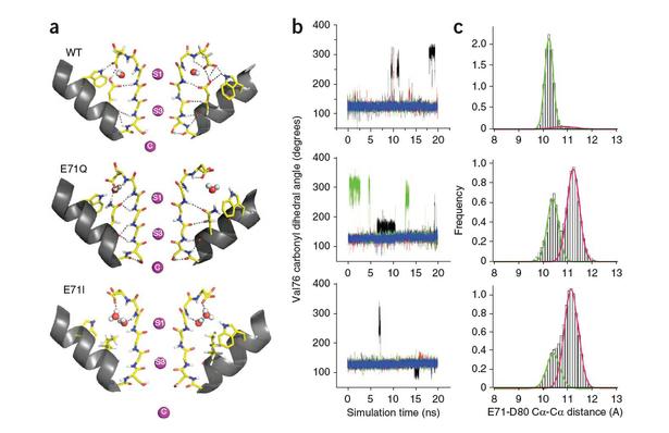
Underlying conformational dynamics of the selectivity filter and the fast gating kinetics. (a) Structural snapshots of outward-facing carbonyl conformations. (b) Dynamics of carbonyl reorientation in KcsA. Time traces of the Val76 carbonyl dihedral angle (N-CA-C-O) during 20-ns molecular dynamics trajectories. Different colored lines correspond to different subunits. Potassium ions were initially placed in the cavity and sites S1 and S3. (c) Distribution of Glu71-Asp80 Cα-Cα distances. The green and magenta fits correspond to populations with Asp80 facing down (centered at ~ 10.3 Å) and ‘flipped’ outward (centered at ~ 11.1 Å), respectively.
Abstract
Modal-gating shifts represent an effective regulatory mechanism by which ion channels control the extent and time course of ionic fluxes. Under steady-state conditions, the K(+) channel KcsA shows three distinct gating modes, high-P(o), low-P(o) and a high-frequency flicker mode, each with about an order of magnitude difference in their mean open times. Here we show that in the absence of C-type inactivation, mutations at the pore-helix position Glu71 unmask a series of kinetically distinct modes of gating in a side chain-specific way. These gating modes mirror those seen in wild-type channels and suggest that specific interactions in the side chain network surrounding the selectivity filter, in concert with ion occupancy, alter the relative stability of pre-existing conformational states of the pore. The present results highlight the key role of the selectivity filter in regulating modal gating behavior in K(+) channels.

