Structural basis of lipid-driven conformational transitions in the KvAP voltage-sensing domain
By Qufei Li, Sherry Wanderling, Pornthep Sompornpisut & Eduardo Perozo.
Published in Nature Structural & Molecular Biology on January 12, 2014. E-Publication ahead of print. PMID: 24413055. Link to publication page.
Core Facility: Membrane Protein Expression/Purification
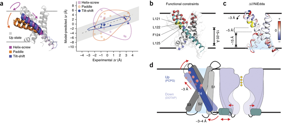
Figure 5. The tilt-shift model. (a) Evaluation of the distance changes, from up state to down state, derived from different gating models in the KvAP VSD. Left, ribbon representation of the up-state VSD model (gray) after MD equilibration and overlapped with putative models based on the helix-screw (magenta), paddle (orange) and tilt-shift (blue) gating mechanisms. Right, expected distance changes against experimental distance differences for the three down states. Gray region indicates the uncertainty of measurement. (b) An alternative explanation for the existing KvAP biotin-streptavidin trapping experiments. All the established state-dependent accessibilities on KvAP focused on the region of 121–125 (in yellow). The paddle model predicts a 15-Å to 20-Å downward S4 movement to account for the streptavidin trapping data (accessibility from the bottom is indicated). (c) ΔΠNiEdda mapped on to the crystal structure, showing water penetration deep into the bottom crevice, where the region 121–125 is only ∼5 Å above the uppermost NiEdda penetration. (d) Cartoon representation of the tilt-shift model on KvAP VSD. The ∼25° tilt and ∼2-Å downward shift of S4 could generate a movement of ∼3–4 Å of the S4-S5 linker, sufficient to open the pore.
Abstract
Voltage-gated ion channels respond to transmembrane electric fields through reorientations of the positively charged S4 helix within the voltage-sensing domain (VSD). Despite a wealth of structural and functional data, the details of this conformational change remain controversial. Recent electrophysiological evidence showed that equilibrium between the resting (‘down’) and activated (‘up’) conformations of the KvAP VSD from Aeropyrum pernix can be biased through reconstitution in lipids with or without phosphate groups. We investigated the structural transition between these functional states, using site-directed spin-labeling and EPR spectroscopic methods. Solvent accessibility and interhelical distance determinations suggest that KvAP gates through S4 movements involving an ∼3-Å upward tilt and simultaneous ∼2-Å axial shift. This motion leads to large accessibly changes in the intracellular water-filled crevice and supports a new model of gating that combines structural rearrangements and electric-field remodeling.
 Right shift of voltage dependence of KvAP in the presence of nonphosphate lipids (80% DOTAP with 20% POPE/POPG at 3:1 ratio (black, Vh = 73 mV, z = 1.5)) compared to the phosphate lipids (100% POPE/POPG 3:1 (red, Vh = −42 mV, z = 3.1)). (b) Schematic of spin labels introduced at positions 16–147 on KvAP VSD one at a time for EPR spectroscopy studies in both phosphate lipid POPC/POPG 3:1 (PCPG) and nonphosphate lipid DOTAP. (c) Mobility (ΔHo−1), oxygen accessibility (ΠO2) and NiEDDA accessibility (ΠNi) of KvAP VSD in PCPG (red) and DOTAP (black). Repeats for 20 selected positions (n = 3–5) are shown. Error bars, s.d. The gray regions represent the four putative transmembrane segments (S1, S2, S3 and S4) from the crystal structure. To facilitate comparison between the two conditions, the area between the two data sets for accessibility profiles for both O2 and NiEdda are colored to highlight the degree and direction of the accessibility changes. With the PCPG data (up conformation) as reference, accessibility increases in the DOTAP-reconstituted sensor are colored yellow, whereas accessibility reductions are colored light blue. Blue and yellow arrows indicate the decreasing key regions that display accessibility decreases or increases, respectively.")
 Changes in water penetration leading to a refocusing of the transmembrane electric field. NiEdda data are mapped onto the crystal structure of the isolated KvAP-VSD for DOTAP (left) and PCPG (center). The color spectrum is a linear scale between white (lowest NiEdda accessibility) and blue (highest). The red spheres depict the lowest two putative gating charges R133 (R6) and K136 (K7) in KvAP. Right, representation of the expected voltage drop along KvAP-VSD after reconstitution in DOTAP (red) or PCPG (black). (b) Differences in ΔHo−1,ΠO2 and ΠNiEdda (DOTAP – PCPG) plotted for the S4 region. (c) Data in b mapped onto the crystal structure of KvAP VSD (PDB 1ORS). ΠNiEdda values increase at the bottom crevice and slightly increase at the top crevice; their values decrease at the S3-S4 loop. ΠO2 increases at the top of S4 (yellow arrow on ΔΠO2 map).")
 Left, scheme of the bifunctional spin label. Right, each distance between a pair of spin labels requires the introduction of four designed cysteine residues. (b) Left, representative raw DEER data for a pair of distance measurements between bifunctional spin label attaching to position 57 and 61 (57/61) and 118/121. Right, distance distribution from Tikhonov regularization. (c) Schematic disagreement in absolute distances of ten pairs in PCPG with the crystal structure, indicating a tilt of the crystal structure by ∼5–8 Å more than in PCPG. (d) Distance differences (DOTAP – PCPG) suggesting a slight tilt and downward movement of S4 at ∼2–3 Å. The distance change from PCPG to DOTAP is consistent in the direction of tilt of the S4.")
