Structure of the Get3 targeting factor in complex with its membrane protein cargo
By Agnieszka Mateja, Marcin Paduch, Hsin-Yang Chang, Anna Szydlowska, Anthony A. Kossiakoff, Ramanujan S. Hegde, and Robert J. Keenan.
Published in Science. 2015 Mar 6;347(6226):1152-5. PMID: 25745174. PMCID: 4413028. Link to publication page.
Core Facility: Synthetic Antigen Binder (SAB) Generation and Crystallography
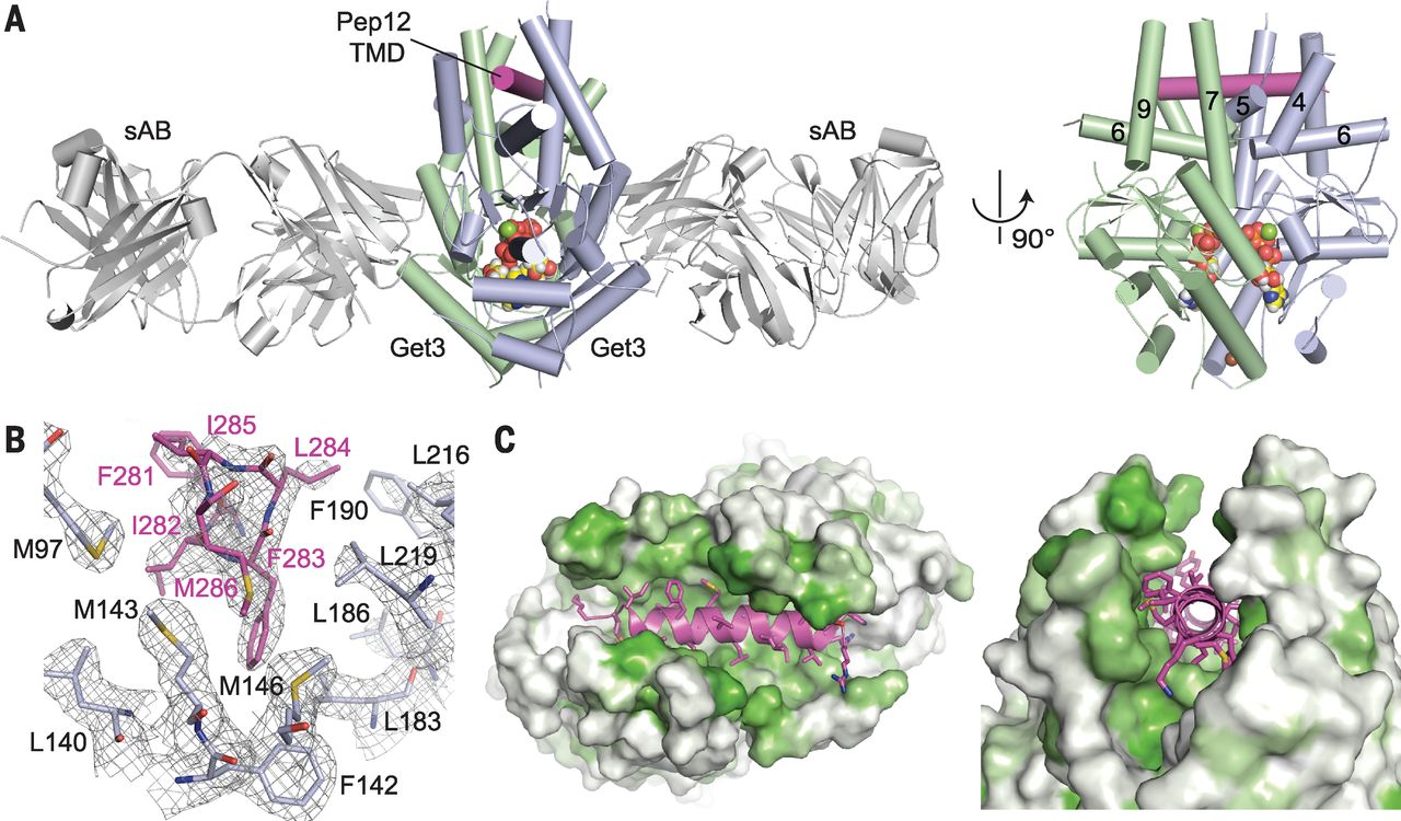
Figure 2. The helical TMD of a TA substrate binds deep within the composite hydrophobic groove of dimeric Get3. (A) Overview of dimeric Saccharomyces cerevisiae Get3 bound to a truncated Pep12 TA substrate (magenta) and nucleotide (spheres), and sandwiched between two copies of an engineered sAB (gray). At right, a “side” view of the complex is shown with sABs removed for clarity. (B) Details of the interaction between the Pep12 TMD C terminus and a methionine-rich cluster at one end of the hydrophobic groove. Electron density is from a 2.05 Å 2Fo – Fc map contoured at 1.0σ. (Amino acid abbreviations: F, Phe; I, Ile; L, Leu; and M, Met.) (C) Surface representations of the TA substrate-binding site, colored from least (white) to most (green) hydrophobic.
Abstract
Tail-anchored (TA) proteins are a physiologically important class of membrane proteins targeted to the endoplasmic reticulum by the conserved guided-entry of TA proteins (GET) pathway. During transit, their hydrophobic transmembrane domains (TMDs) are chaperoned by the cytosolic targeting factor Get3, but the molecular nature of the functional Get3-TA protein targeting complex remains unknown. We reconstituted the physiologic assembly pathway for a functional targeting complex and showed that it comprises a TA protein bound to a Get3 homodimer. Crystal structures of Get3 bound to different TA proteins showed an α-helical TMD occupying a hydrophobic groove that spans the Get3 homodimer. Our data elucidate the mechanism of TA protein recognition and shielding by Get3 and suggest general principles of hydrophobic domain chaperoning by cellular targeting factors.
Editor’s Summary
How to GET to the right membrane
Membrane proteins with a hydrophobic transmembrane domain (TMD) play critical roles in virtually all aspects of cell physiology. After it has been synthesized in the cytosol, this TMD must be targeted to and inserted into the correct membrane. The GET pathway is one of two targeting pathways to the endoplasmic reticulum conserved across all eukaryotes. It is not clear how the central targeting factor, Get3, recognizes a TMD to shield it from aggregation until it is successfully inserted into the membrane. Now, Mateja et al. show that the functional targeting complex comprises a Get3 dimer bound to a single TMD. The helical hydrophobic TMD binds deep within a large hydrophobic groove in the Get3 dimer. This groove closes slightly upon TMD binding, forming a dynamic “lid” over the mouth of the groove.
 Experimental strategy. (B) SGTA-TA or Get3-TA complexes (figs. S1 and S3) at 1 μM were incubated with 1 μM of the indicated proteins, followed by amine-reactive cross-linking. Reactions were analyzed by SDS–polyacrylamide gel electrophoresis and Coomassie blue staining to detect the input proteins (top) or autoradiography to detect the 35S-labeled TA protein cross-links (bottom). (C) Reactions as in (B) were monitored by sulfhydryl-reactive cross-linking for TA protein release from SGTA (bottom). Reactions contained 0.5 μM of each factor, except lanes 4 and 5, which contained Get4-Get5 at 0.1 and 0.2 μM, respectively. Asterisks next to Get3 or SGTA indicate point mutants that disrupt interactions with Get4 or Get5, respectively. (D) Products of the indicated transfer reactions were incubated with yeast rough microsomes (yRM) and analyzed for insertion. (E) Sucrose gradient size analysis of Get3-TA complex formed by Get4-Get5–dependent loading from SGTA (red). Free, dimeric Get3 (gray) and E. coli–produced tetrameric Get3-TA substrate complex (black) are shown for comparison (fig. S5). Peak fractions containing substrate (red) were analyzed directly or after cross-linking and immunoprecipitation for Get3 to specifically detect Get3-TA complexes (bottom panel).")
 SGTA-TA complexes were prepared and subjected to transfer reactions with wild-type (WT) and mutant Get3 proteins as in Fig. 1C. LL-SS (L183S/L186S), LLL-SSS (L183S/L186S/L219S), and FL-DD (F190D/L216D) are hydrophobic groove mutants; E253R is a mutation that disrupts interaction with Get4. (Amino acid abbreviations: D, Asp; E, Glu; F, Phe; L, Leu; R, Arg; and S, Ser.) (B) “Top” and “side” views of Pep12 (magenta), Nyv1 (blue), and Sec22 (green) complexes superimposed on the free Get3 closed dimer structure (yellow; PDB code: 2woj). Relative to free Get3, the end of helix 7 extends and begins to curve inward over the substrate. (C) WT or benzophenone-containing (at the indicated positions) Get3-TA complexes were prepared as in fig. S3, and the dimer peak was subjected to ultraviolet (UV) cross-linking. Uncrosslinked TA protein and its adducts to one or two Get3 proteins are indicated. (D) “Side” views of the Get3 dimer, looking into the groove. In its transient empty state, Get3 is splayed apart, with two hydrophobic “half-sites” occupied by the helix 8 region. ATP binding drives Get3 into a closed conformation, which is captured by two copies of the Get4-Get5 complex. In this state, helix 8 is displaced, and the composite hydrophobic groove is now preorganized for substrate binding. After substrate transfer from Sgt2, the targeting complex is released. The helix 8 region now dynamically shields the substrate during transit to the ER membrane.")
