Winner of the logo design contest announced

In July of this year, we announced a Consortium logo design contest. The contest was open to all post docs, graduate students, lab technicians, and lab personnel associated with the Consortium, who were asked to submit a stylized design that informs and captures the spirit of the Consortium. These logos were reviewed by the Executive Committee of the MPSDC, who judged the logos based on creativity, originality, aesthetics, use of space, and color. The prize for the winning logo designer was an Apple iPad with Wi-Fi and 32GB Memory. To our delight, we received over a dozen entries from a number of affiliate laboratories. Some of the entries that were submitted can be viewed here.

Upon much deliberation, we decided upon two finalist submissions: one of the logos submitted by David Medovoy from the Perozo and Roux labs, and the logo submitted jointly by Amelia Randich from the Perozo lab and Ernesto Vargas from the Roux lab. We asked the designers to revise their logos in accordance to some of our criteria, and this is what they came up with:


By David Medovoy


By Amelia Randich and Ernesto Vargas

In the end, we decided to select David Medovoy’s logo as the winner, which is now on display on the top left of the website. David goes home with the coveted prize, but we gave Amelia and Ernesto an iPod shuffle for their efforts as well.

We would like to thank all participants for their creativity and hard work!

MPSDC launches new website

We are delighted to announce the launch of the newly redesigned Membrane Protein Structural Dynamics Consortium (MPSDC) website. This new website is intended to be a gateway to the Consortium’s research and publications as before, but also a means for us to reach out to and engage with the scientific community and the public at large. We have also made a number of tweaks that we hope will improve the user’s browsing experience.

We invite you to take a look at the following new or refurbished sections and pages:

  • The new Homepage, which presents all of the latest updates from the MPSDC and is intended to be a guide to the website at large.
  • Mission Statement, providing a succinct and current overview of the Consortium’s goals and ambitions.
  • A page dedicated to our new initiative aimed at maximizing the reach and investment of the MPSDC through direct or indirect interactions with the scientific community at large: Associate Members. To find out more about associate members and a current list of AMs, please visit this page.
  • The Community section is entirely new, and much of the effort in giving our research a broader purview and significance can be found here. We provide external links to related scientific initiatives, and access to various educational resources that we’ve created. Importantly, we offer Consortium affiliates the opportunity and forum to reflect on their research, as well as the field at large. Finally, we’ve created a subsection that will allow Consortium affiliates to write their own guide to the field, under the heading Ideas and Concepts. There is much more to come within this section so stay tuned.

In addition, we have incorporated a new logo for the Consortium, which was designed by David Medovoy from the Roux and Perozo labs. David’s logo was selected as the winner of the Logo design content, and has graciously tweaked his original design to fit the theme of the website. Congratulations, David!

We’ve made many other changes and additions throughout, and there is still more to come. Overall, we hope that these changes will provide an enhanced perspective of the Consortium’s collaborative activities and augment our communications with the scientific community. We would love to hear from you and welcome any suggestions, corrections, comments, or compliments. Let us know what you think by sending us an e-mail or in the comments section below!

Worth One’s Weight

By Raymond E. Hulse
Department of Biochemistry and Molecular Biology, The University of Chicago

It is an understatement to say that the structural solution of the highly sought after voltage gated sodium channel is worth its weight in salt. Derived from a voltage gated bacterial sodium channel the solution, from Arcobacter butzleri (NaVAb), required a creative and demanding approach. The authors used techniques to achieve this structure that differ from other bacterial ion channels such as insect cell expression, the use of digitonin to solubilize and stabilize the protein, a lipid/detergent system for crystallization and strategic cysteine mutagenesis among several techniques. This achievement has been highly sought after since the discovery of bacterial voltage gated sodium channels and many researchers have spent considerable effort and time towards its elucidation.
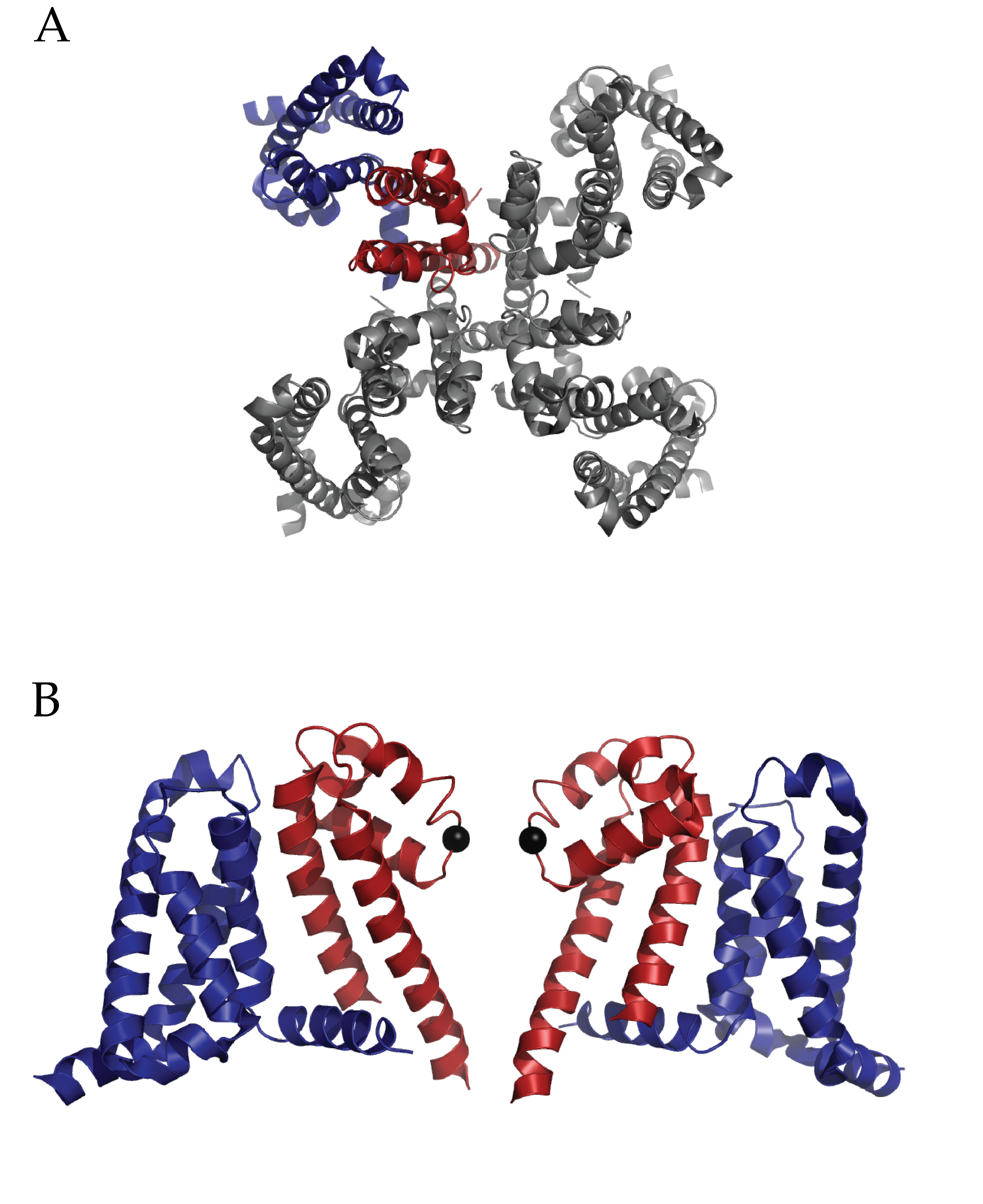
Figure 1 (click to enlarge)

Structure of Full length NaVAb: A) Extracellular top view of NaVAb. In dark blue is the VSD and in dark red is the PD of one monomer. B) A side view of two of four monomers (removed to ease visualization) of NaVAb. The alpha carbon of Glutamate 177 is represented as a black sphere. (PDB ID 3RVY)

Payandeh et al’s work will invigorate research into such critical questions as how Na+ ions are coordinated at the selectivity filter, alternative conformational states of a voltage sensor and the mechanism of voltage-dependent drug block via local anesthetics1. The pore domain of NaVAb is unique in that is possesses a helix-loop-helix conformation which deviates from well-researched potassium channels. The voltage sensor domain (VSD) also presents a surprise; the entire charge-sensing segment, S4, has an atypical secondary conformation of a 3-10 helix. Finally, side fenestrations in the central cavity of the pore domain are also present and provide a unique opportunity for further hypothesis driven research into pharmacological research. Notably the authors interpret their findings of this structure as a pre-activated voltage sensor with a closed pore.

One critical question surrounding this structure is the nature of coordination of Na+ ions. While potassium ion channels utilize the ability of the selectivity filter to dehydrate K+ ions, the Na+ ion is either incompletely dehydrated or not at all. The selectivity filter of voltage gated bacterial sodium channels is highly conserved. This structure demonstrates the role of those conserved sequences by revealing an intricate scaffold of hydrogen bond interactions between the selectivity filter and the two adjacent pore helices. Interestingly, these interactions occur within and between monomers. The structure does implicate the importance of one particular residue in the selectivity filter, a glutamate. The carboxylate group shares hydrogen bonds with nearby backbone amides on pore helices and the entire side chain forms the narrowest part of the pore. Yet there were no observed Na+ ions in the pore domain of the structure, which the authors ascribe to a low affinity.

In what is already a lively field of study, the conformational state of the VSD of NaVAb is striking. Foremost, the presence of a 3-10 helix along the sensing charges (R1-R4) in S4 is unique among all VSDs observed to date.  Typically, a complete to partial alpha helix has been observed. An extensive network of interactions through the entire VSD is present.  These include well-researched electrostatic and hydrophobic interactions with the charge sensor S4 in addition several structure specific types among the three other alpha helices (S1, S2 and S3). The authors interpret the structural information of the voltage sensing domain, in conjunction with previous disulfide locking experiments and comparison to other structures as evidence for an activated sensor. This is a remarkable finding considering the protein is not in the presence of an electric field and so not stimulated. Is it possible that the structure has trapped a different conformation such as an inactivated state? Functional assays on the particular cysteine mutations used to crystallize this structure may help further understanding.
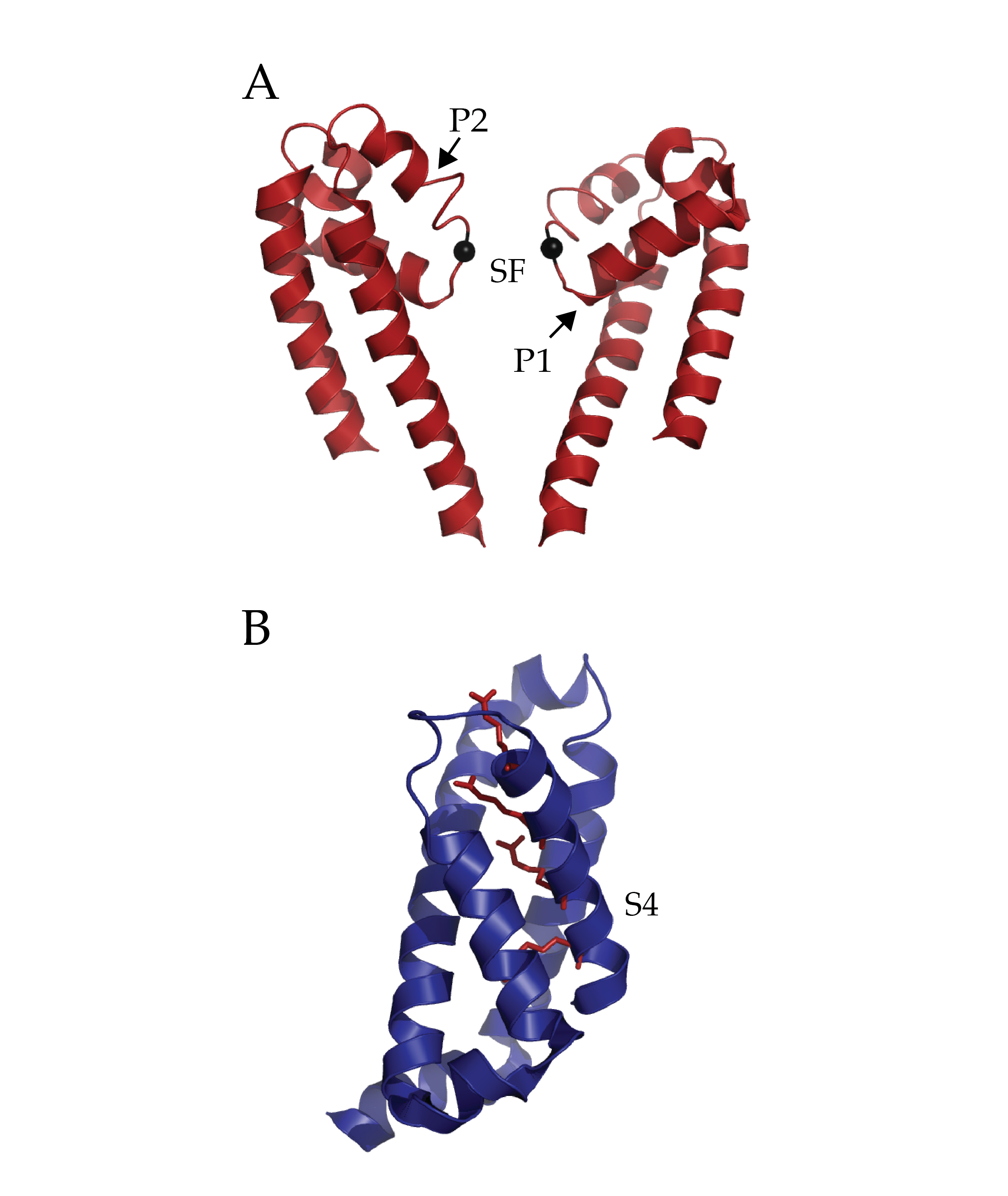
Figure 1 (click to enlarge)

Isolated Pore and Voltage Sensing Domain of NaVAb. A) Two of four pore domains isolated from remaining structure. The two pore helices are indicated with arrows (P1 and P2) and the selectivity filter (SF). The alpha-carbon of glutamate 177 in the selectivity filter has been rendered as a black sphere to ease localization. B) A single voltage sensing domain of NaVAb is depicted in cartoon format with the four sensing charges (R1-R4) of the S4 helix depicted in stick format (dark red). (PDB ID 3RVY)

Voltage gated sodium channels have long been researched in the context of pharmacology. This includes understanding the mechanism of pharmacological agents such as antiarrhythmics and local anesthetics (e.g., lidocaine). The side fenestrations of NaVAb are notable for their location; within the central cavity of the pore-domain itself. The presence of these windows gives the author an opportunity to present an interesting hypothesis: Are these the sites of use-dependent anesthesia block of sodium channels?  The local hydrophobic environment might present a route for such drugs (e.g., etidocaine) to partition to the core of the membrane and then enter into the fenestration. The presence of a conserved phenylalanine residue at the fenestration with nearby conserved residues implicated in binding in eukaryotic channels is tantalizing. Ironically, while this family of bacterial voltage gated sodium channels select for Na+, they can be blocked by dihydropyridines (which block L-type Ca2+ channels). Future research into these mechanisms of pharmacological action will be enhanced greatly by this and future structures.

There are many interesting questions that arise from this work. How well does this particular channel represent both prokaryotic and eukaryotic voltage gated sodium channels? Bacterial voltage gated sodium channels are homotetrameric and so possess symmetry. Eukaryotic voltage gated sodium channels have a substantially more complex topology of a single, asymmetrical protein with 24 trans membrane helices.  Additionally, kinetics of activation and inactivation differ between the two classes of channels. Sequence comparison of the growing family of bacterial voltage gated sodium channels to this new channel reveal several interesting differences. NaVAb lacks two glycines which have been noted for their role in gating for other bacterial voltage gated sodium channels2-4. NaVAb also appears to inactivate rapidly relative to other bacterial voltage gated sodium channels. Could this characteristic arise from minor sequence variations in the pore domain suggesting a potentially different mechanism of c-type inactivation or gating in this channel as has been observed in NaChBAC5? Finally how would mutations performed in earlier voltage-gated sodium channels in the selectivity filter promote Ca2+ selectivity6? Would such mutations alter the scaffold in the pore domain and change its stability and mechanism for selection?

Truly it is an exciting time for the sodium channel community in specific and the ion channel community in general.  This work has great potential to help these communities understand the mechanism of how sodium channels work but also serve as a stepping stone towards understanding the nature of Ca2+ selectivity, perhaps the only topic more interesting than Na+ or K+ selectivity.

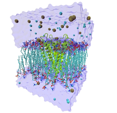
The structure of NaVAb has been rendered in cylinder format to facilitate visualization of the entire structure and rotated along the selectivity filter’s axis of symmetry. (PDB ID 3RVY)


Citations

1.     Payandeh, J., Scheuer, T., Zheng, N. & Catterall, W.A. The crystal structure of a voltage-gated sodium channel. Nature 475, 353–358 (2011).

2.     Irie, K. et al. Comparative study of the gating motif and C-type inactivation in prokaryotic voltage-gated sodium channels. J Biol Chem 285, 3685–3694 (2010).

3.     Zhao, Y., Scheuer, T. & Catterall, W.A. Reversed voltage-dependent gating of a bacterial sodium channel with proline substitutions in the S6 transmembrane segment. Proc Natl Acad Sci USA 101, 17873–17878 (2004).

4.     O’reilly, A.O. et al. G219S mutagenesis as a means of stabilizing conformational flexibility in the bacterial sodium channel NaChBac. Mol Membr Biol 25, 670–676 (2008).

5.     Pavlov, E. et al. The pore, not cytoplasmic domains, underlies inactivation in a prokaryotic sodium channel. Biophys J 89, 232–242 (2005).

6.     Yue, L., Navarro, B., Ren, D., Ramos, A. & Clapham, D. The Cation Selectivity Filter of the Bacterial Channel, NaChBac. Journal of General Physiology 1–9 (2002).
[/EXPAND]

Site-directed spin labeling: Conformational dynamics in proteins

By Eduardo Perozo and Hassane Mchaourab

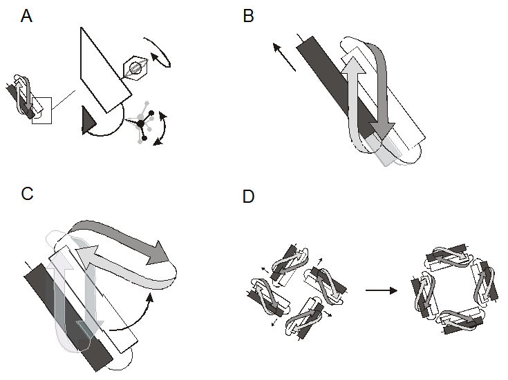
The functional behavior of most proteins is intimately related to their conformational dynamics. It follows that a thorough understanding of structure-function relations in a given protein requires a molecular description of the types and extent of the protein movements underlying function. This includes catalysis, transport, cellular locomotion, regulation of activity, and formation of protein assemblies. X-ray and NMR structures have made it clear that proteins undergo a wide variety of motions, whether as a consequence of a triggered action (ligand binding, protein-protein contact, physical stimuli) or as the result of the natural fluctuations around a flexible linker between domains. Closer examination reveals that the types and dynamic behavior of these movements can be hierarchically divided according to four major groups (Figure 1): Side-chain rotamers (ps-ns), reorientation of secondary structure elements (ns), large domain movements (ns-ms), and whole subunit rearrangements in cooperative oligomeric complexes (ms-ms).

Figure 1 (click to enlarge)

The types of molecular motions in proteins. A. Side-chain rotamers, B. Reorientation of secondary structure elements, C. Large domain motions, D. Whole subunit rearrangements.

Among the types of mechanisms of motion mentioned above, by far, the best understood is that occurring among protein domains. Gerstein and co-workers have reviewed and classified domain motions from a growing database of proteins with known three-dimensional structures (Gerstein et al., 1994). They concluded that most domain motions ultimately originate from two basic mechanisms, hinge bending or shear motions. In hinge bending, motion occurs due to changes in a few main-chain torsion angles along a localized region of the protein connecting two interacting modules (strands, b-sheets, a-helices, or combinations of them). These two modules are normally not constrained by tertiary packing interactions, and the transition from “open” to “closed” positions is accompanied by the exclusion of water molecules from the inter-domain space. In contrast, interactions among tertiary structure elements are severely constrained due to packing interactions, precluding large changes in main-chain torsion angles. Hence, shear motions have been observed mostly among closely packed regions of proteins. Shear involves small changes in side-chain torsion angles among interdigitating side-chains, without large changes in main chain conformation. These can be observed either parallel or perpendicular to the interface of closely packed segments of a polypeptide chain. An argument is also made that because these types of protein rearrangements are usually fast, transition between the two conformations cannot involve large energy barriers. This is particularly relevant in the analysis of equilibrium fluctuations, since the relatively weak forces involved in crystal packing are known to favor one of the two possible conformations, as in the case of the T4 lysozyme (see below). A number of useful databases of protein motions continue to be maintained on the web by Gerstein and co-workers and by Hayward and coworkers.

It can be argued that one of the most powerful aspects of site-directed spin labeling as a structural approach, is its potential to discern the types of protein motions described above. Information on the relative movement of secondary structure elements, domains or whole subunits can be obtained by studying patterns of change in probe mobility and solvent accessibility or changes in relative distance among residue pairs between domains or subunits. However, because this is a reporter group technique, side-chain rotamer conformations cannot be analyzed.


Animation courtesy of Dr. Peter Fajer, Florida State University.

Analysis of equilibrium fluctuations

Protein conformational equilibria are the results of the thermally activated interconversion of the structure between multiple states of similar energies. These motions differ in their amplitude, time scale and biological consequence. Thermally activated equilibrium fluctuations in the structure can facilitate access of a substrate to an active site cleft, binding of an effector to an allosteric site and transport of ions through channels. Because thermally activated equilibria involve states of similar energies, lattice forces may be sufficiently large to select a particular state in a crystal, obscuring the existence of others. In some cases, crystallization of the same protein in two different crystal forms allows the observation of multiple conformations. Dynamics can also be inferred from the occurrence of disorder in the crystal and analysis of thermal factors.

Conformational equilibria in solution can be explored by SDSL as long as the interconversion rate is within the time scale of the employed EPR technique. Since the dynamic range of EPR covers over 7 orders of magnitude, it provides a suitable window for many protein motions of interest. Both the mobility of the nitroxide side chain and the distance between the nitroxide and a second paramagnetic center can be used to detect conformational changes. The structural determinants of these observables as well as their resolution makes them ideal to detect large scale concerted motion of large segments of the protein molecule. The population of multiple structures results in a heterogeneous environment in the vicinity of the nitroxide that is reflected in the EPR spectral lineshape as multiple motional states. Similarly, if the dynamics involve rigid body motion of a secondary structural element it will modulate the proximity of an attached nitroxide to a nearby reference paramagnetic center. Although the contribution of the backbone flexibility to the mobility of the nitroxide is not well understood, in principle backbone fluctuations are also detectable. Because conformational equilibria involve motions that are constantly occurring in solutions, the spectral properties are ensemble averages with each state with distinct spectral characteristics contributing to a different degree.

In practice, detailed quantitative interpretation of EPR data arising from such ensembles are yet to be developed. At the fundamental level, multicomponent EPR spectra can arise from different conformations of the spin label itself. Furthermore, the addition of parameters such as the number of intermediate states and the mobility and/or internitroxide distance in each state makes the linear CW-EPR spectrum underdetermined.

The problem is considerably simplified if the structure of one of the conformers is known. This provides a reference state against which spectral observables can be compared. An ideal situation arises if the states of known structure can be selectively populated via changes in the physicochemical conditions or by binding of a ligand or a substrate. In this case, reference EPR spectra can be obtained and the sign of the changes in mobility or interresidue distances when multiple states are populated can be interpreted in terms of the nature, extent and magnitude of the movement involved in the equilibria.

Online Workshop on Simulating Membrane Channels hosted by Emad Tajkhorshid

Computer Simulation of Biological and Artificial Membrane Channels

Modeling and simulation of molecular systems have become inseparable components of modern research in molecular biology. In particular, molecular dynamics simulation is increasingly used to investigate the mechanism and structure-function relationship of a wide range of biological macromolecules. The course will present a brief introduction to such methodologies, and uses membrane proteins as an example of how computational biophysical methods and simulation technologies can be applied to biological problems. The molecular systems used as examples will include biological water channels, as well as carbon nanotubes as artificial materials used in nanotechnology. The goal of the course is to walk the participants through various steps involved in modeling, simulation setup, and analysis of the results of molecular dynamics simulations and discuss what can be learnt from simulation studies of these systems. The lectures are accompanied by hands-on tutorials in which the students can set up and run simulations on their own.

This workshop, hosted by Emad Tajkhorshid, will take place on November 28 – December 1, 2011. It is designed for graduate students and postdoctoral researchers in computational and biophysical areas. Working familiarity with the VMD and NAMD software used in the workshop is assumed. Streaming video of a recorded lecture, tutorial self-study, and Skype/phone contact with the instructor are used to deliver course content. Enrollment is limited to ten participants. There is no fee for the workshop.

Find out more information and apply »

Categories
Recent Posts
Archives
Recent Comments
Recent Photos