New SNPS (Symmetric Nano-Positioning System) software made available

Proteins may undergo multiple conformational changes required for their function. One strategy used to estimate target site positions in unknown structural conformations involves single-pair resonance energy transfer (RET) distance measurements. However, interpretation of inter-residue distances is difficult when applied to 3D structural rearrangements, especially in homomeric systems. Probe diffusion further complicates this task by biasing measurements towards shorter distances. Lanthanide resonance energy transfer (LRET) is an ideal technique for simultaneously resolving multiple distances within a protein. Hyde et al. (2012) recently combined LRET with an ensemble of numerical methods to form the Symmetric Nano-Positioning System (SNPS), which allows accurate 3D positioning and inter-probe distance estimation in functional homomeric proteins.
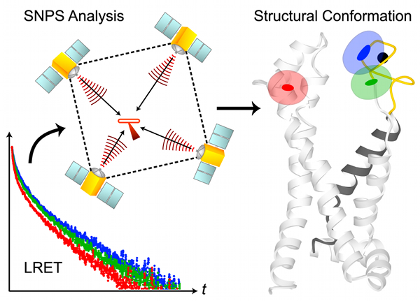
SNPS determines the 3D position of lanthanide donors [satellites] attached to a target site (one per subunit), relative to a single fluorescent acceptor [antenna] placed in a static reference site, as illustrated in the above figure. The acceptor’s position and accessible volume can be modeled from its structure-based labeling site by several methods, including dihedral scan analysis. SNPS can be applied to all defined conformational states of the protein and with simultaneous functional recordings. Satellite-to-antenna distances are encoded in time-resolved LRET lifetime decays. SNPS directly fits a 3D geometric model of satellite positions to LRET lifetime measurements using an inverse trilateration-based curve fitting procedure. Global analysis implementation fits the geometric model to an ensemble of replicate measurements. Numerical and analytical tools are integrated to account for probe diffusion and evaluate the confidence region of fitted positions. SNPS is well-suited to estimate 3D conformational changes at the target site between defined conformational states. In its first application, SNPS was used to determine the position of a functional voltage-gated potassium channel’s voltage sensor in its three major conformations [H. Clark Hyde, Walter Sandtner, Ernesto Vargas, Alper T. Dagcan, Janice L. Robertson, Benoit Roux, Ana M. Correa, Francisco Bezanilla (2012), Structure 20(10): 1629-1640].

The SNPS Toolbox contains the following stand-alone programs that implement the SNPS method:

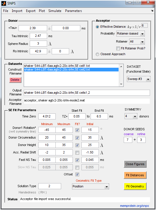
  • SNPS: Performs inverse trilateration-based curve fitting of LRET lifetime decays to estimate the 3D position of lanthanide donors [satellites] attached to a target site (one per subunit), relative to a single fluorescent acceptor [antenna] placed in a static reference site. An acceptor file must be supplied by the user that specifies the acceptor’s coordinates and accessible volume. SNPS can alternatively fit all donor-acceptor distances constrained such that donors must lie in a polygon (symmetric) geometry, without any knowledge of acceptor position. SNPS can also simulate LRET lifetime decays at a user-defined donor position and noise level to explore the relation between donor position and decay shape. LRET lifetime decays are expected to be sensitized emission measured by a photomultiplier tube in analog mode (Poisson noise). The user imports lifetime decay files (ASCII .txt) and an acceptor file (ASCII .txt or Matlab .mat). See the “Example Data” folder for examples of required formatting. Fits are accompanied by a comprehensive set of output figures and a solution file.
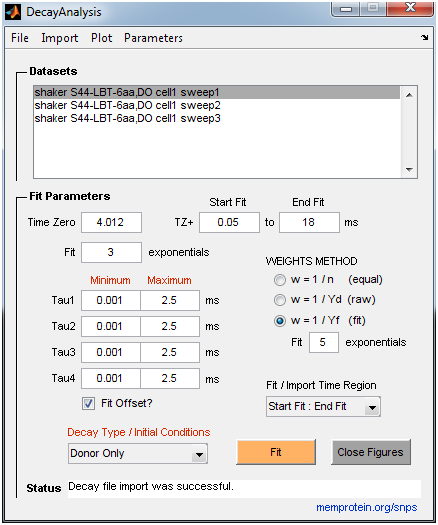
  • DecayAnalysis: Performs model-free curve fitting of multi-exponential decays to estimate time constants and amplitudes (up to 4 exponential components). It is intended for donor-only time constant analysis of LRET experiments, but can also be used for general exponential fitting. The donor-only time constant is a required parameter for the SNPS program. The user can place constraints on time constants and define a weights scheme appropriate for the measurement uncertainty (e.g., Poisson vs. Gaussian noise). A large set of measurements can be fit quickly with basic statistical analysis reported. The user imports lifetime decay files in ASCII (.txt) format. See the “Example Data” folder for examples of required formatting.

SNPS and Decay Analysis Screen Guides


Download the software (version 2012.1):
Windows 64-bit: SNPS_Toolbox_win64.exe
Windows 32-bit: SNPS_Toolbox_win32.exe

Installation: Clicking on the downloadable .exe file will directly launch the Matlab Component Runtime (MCR) installation process and extract the SNPS and DecayAnalysis applications. This is a one-time installation, after which both applications can be launched. Installation of the included MCR is required even if you currently have Matlab or other MCR versions installed. Regarding processing speed, CPU speed takes precedence over the number of cores. The extracted folder also contains an “Example Data” folder with example data from the publication: AgTx2[II]-D20C-BODIPY-FL-maleimide acceptor cloud and Shaker S4(4) LBT construct sensitized emission (SE) and donor-only (DO) lifetime decays. Fit progress and information is displayed in an accompanying DOS window. Output files are written to a subfolder of the imported data directory.

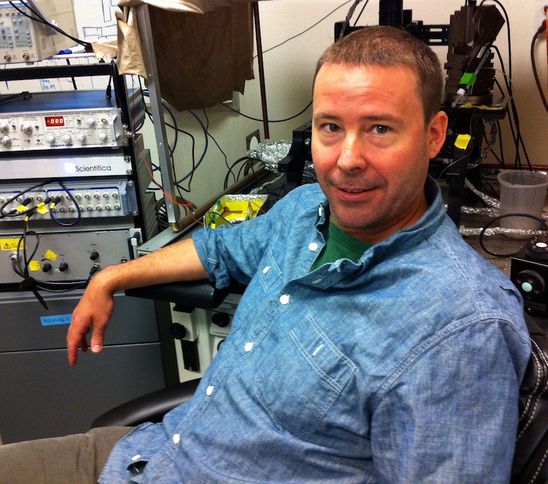
The software was developed and written by H. Clark Hyde from Francisco Bezanilla’s group at the University of Chicago. Please contact us with questions or suggestions using the comments form below.

MPSDC welcomes Wonpil Im and Chris Ahern!

MPSDC is pleased to welcome two new members to the team!

Chris Ahern, University of British Columbia. Click to enlarge.

Christ Ahern joins us from the University of British Columbia where he works as an associate professor in the Department of Anesthesiology, Pharmacology and Therapeutics.

Wonpil Im, The University of Kansas. Click to enlarge.

Wonpil Im will be working with the consortium from The University of Kansas, where he works as an associate professor in the Department of Molecular Biosciences.

We look forward to collaborating with both scientists. They bring unique skills to the consortium and are particularly well qualified to help advance our interdisciplinary research goals. Their contributions will help the consortium continue to gain momentum in our effort to elucidate the relationship between membrane protein structure, function and dynamics.

The next MPSDC annual meeting will be May 9-10, 2013. Save the date!

Save the date for the next annual meeting scheduled to be held May 9-10, 2013. The location will be here in Chicago, and the venue will be announced soon!

In May 2012 the Membrane Protein Structural Dynamics Consortium (MPSDC) held its first Frontiers in Membrane Protein Structural Dynamics conference at the Gleacher Center in Chicigao. This year our annual meeting will provide similar opportunities for group conversation and collaboration between participants. We hope you will join us!

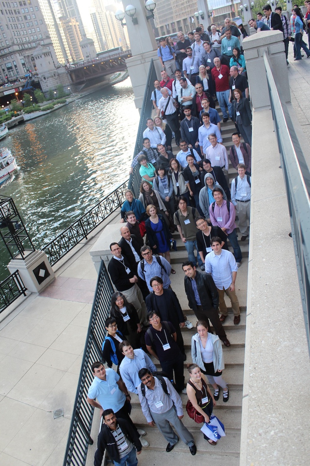
Participants gathered on the stairs at the Gleacher Center for a group photo.

First Frontiers in Membrane Protein Structural Dynamics Conference was a success


Dorothee Kern, Brandeis University and External Advisory Committee Member

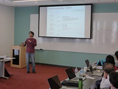
On May 3rd and 4th, the Membrane Protein Structural Dynamics Consortium (MPSDC) held its first Frontiers in Membrane Protein Structural Dynamics conference. The conference consisted of scientific sessions and poster presentations, and featured both Consortium members and external invitees. Attendance was open to the public took place within the context of our 3rd Annual MPSDC Meeting, where all members, NIH representatives and our External Advisory Committee participated. Prior to the conference, the MPSDC’s Computational Modeling Core hosted a NAMD/VMD workshop and a mini-symposium concerning the latest advances in membrane protein modeling.


Miguel Holmgren, NINDS and collaborator in Bridge 1: Conformational Transitions in P-class ATPases

Both the conference and CMC events were very well attended and enabled extensive conversations surrounding the topic of cutting edge advances and scientific methods in the field of membrane protein dynamics, as well as ways to resolve current roadblocks. The conference was able to hit a high note in bringing together the issues and ideas most relevant to the key goals of the consortium both in the present and in the future. Chris Ahern of the University of British Columbia and MPSDC Associate Member noted afterwards that it was probably one of the best meetings he’s been to, “primarily because of the quality of the science that’s being done, as well as the excitement and eagerness of people to cooperate.”


José Faraldo-Gómez, Max Planck Institute for Biophysics and MPSDC Associate Member

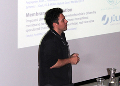
In the afternoon, two discussion panels were held, which themselves are in the spirit of much of the broader conversations that took place at the conference as a whole. The first panel, “Finding a common language: linking experiment and computation” was chaired by Hassane Mchaourab, and included Benoît Roux, Martin Zanni, Ivet Bahar, Dorothee Kern, and Emad Tajkhorshid as participants.


Chris Ahern, University of British Columbia and MPSDC Associate Member

The second panel was titled “Breaking the barriers of membrane protein expression and labeling” and was moderated by Robert Nakamoto. The panel included Chris Ahern, Jim Bowie, Volker Dötsch and Shohei Koide. Discussion focused on three topics: the optimal nitroxide spin probe for monitoring protein dynamics and DEER distance measurements, how to incorporate such a probe into the protein targets, and cell free synthesis of target proteins. The optimal nitroxide spin probe would be connected to the backbone by only a β carbon. Such a label can be introduced using chemical synthesis, but because most of our proteins are too large, methods for bio-incorporation of unnatural amino acid are preferred. Cell free biosynthesis systems such as those developed in the laboratory of Volker Dötsch, Goethe University, and Chris Ahern, University of British Columbia, may provide the best approaches. The Protein Core labs will explore methods for charging the non-sense tRNA by evolved tRNA synthetases or a ribozyme using technologies called the Flexizyme developed by Soga and co-workers at Tokyo University. The acylated TAG tRNA charged with the spin probe is simply added to the cell free synthesis mix. Another issue is the lability of nitoxides to reduction by ascorbic acid. We will test a variety of nitroxide spin probes, which have been reported to be relatively insensitive to reduction. Finally, we discussed specific placement of labels using synthetic binders. In particular, the synthetic 10FN3 binders, or monobodies (~93 aa, derived from a type III fibronectin domain), can mediate specific labeling of a protein and effectively attach a label or cargo to the protein target with high affinity and stability.

We’d like to thank all who attended and partook in the discussions. By all accounts, it is hard to think of a better outcome for the conference and accompanying events, and we look forward to hosting another meeting in two years.

Below is a gallery displaying photos of the conference. You can either scroll through the photos here or visit the set on Flickr. We’ve also made available several brief audio interviews with attendees of the conference, to be found in the margins of the body of this post.

New Continuum-Molecular Dynamics (CTMD) software made available by Computational Modeling Core

The membrane environment is a key determinant of function and/or organization of membrane proteins. It is essential for the activities of the Consortium to have access to quantitative information about the characteristics and energetics of membrane-protein interactions. For multi-segment transmembrane proteins in particular, such characterization is complicated by the different hydrophobic thicknesses of the component transmembrane segments and the radially non-uniform hydrophobic interface created between the membrane and the protein. A hybrid Continuum-Molecular Dynamics (CTMD) approach to compute membrane deformations profiles around multi-segment proteins and corresponding energetics was recently published [Sayan Mondal, George Khelashvili, Jufang Shan, Olaf Andersen, Harel Weinstein (2011), Biophysical J (101): 2092-2101; link]. This stand-alone application implements the CTMD approach.

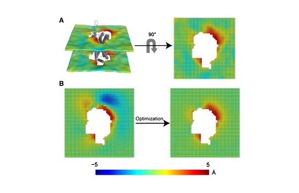
Protocol for the 3D-CTMD approach, illustrated for rhodopsin in a diC14:1PC lipid bilayer (Mondal et al., BJ 2011). Click to enlarge and learn more.

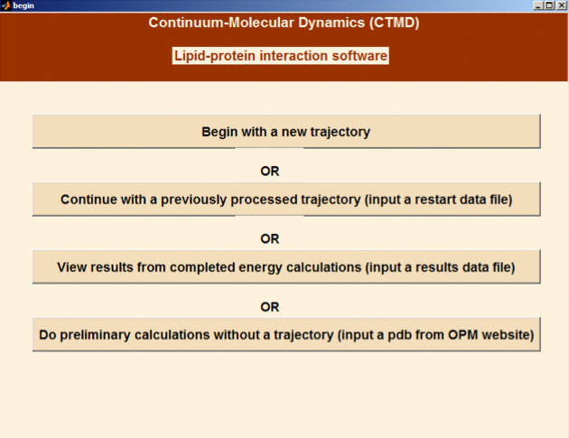
The CTMDapp software calculates the deformation profiles of the bilayer and the free energy cost of the membrane deformation around multi-segment transmembrane proteins, taking into account the radially non-uniform hydrophobic surface of the protein. As the primary input it requires a molecular dynamics trajectory of a multi-segment, transmembrane protein embedded in a bilayer.

To allow for calculations without a molecular dynamics input, the CTMDapp software also implements a simplified version of the CTMD method that can assess the radial asymmetry of the membrane-protein interface for a particular protein structure at an approximate level.

Methodological details can be found in the original paper (Mondal et al., BJ 2011). In addition, the application is documented with brief usage notes at every step and generates diagnostic intermediate output.

Download the software:

Instructions:
For the Windows version, clicking on the downloadable .exe file will directly launch the Matlab Component Runtime (MCR) installation process and extract the app executable. For the Mac version, the MCR must be installed first by opening MCRinstaller.dmg in the folder. This is a one-time installation, after which CTMDapp can be launched. The Unix version requires an interface like Xwin along with a script setting proper paths.

The software was written by Sayan Mondal (mos2013@med.cornell.edu) in the Harel Weinstein (haw2002@med.cornell.edu) lab at Weill Cornell Medical College, Cornell University. Please feel free to contact us with questions and suggestions about the software.

Categories
Recent Posts
Archives
Recent Comments
Recent Photos