Final Program for III Frontiers in Membrane Protein Structural Dynamics Announced

Friends,

As we get everything in order to welcome you this Friday to our Frontiers in Membrane Proteins Structural Dynamics Conference, we wanted to share with you the final program of the meeting.

The next few days promise to be a truly exceptional survey of membrane proteins structure and dynamics today. A veritable state of the art of the field. With our two keynote speakers (Chris Miller and Olga Boudker) leading 8 individual mini-symposia.

Personally, we are very much looking forward to seeing you and interacting with you later this week.

All the best,

The Organizing Committee


III Frontiers in membrane Protein Structural Dynamics
APS Conference Center, Argonne National Lab

Friday, November 10

7:45 Continental Breakfast, APS 402 Atrium
8:15 Eduardo Perozo (University of Chicago), Stephen Streiffer (Advanced Photon Source)
Welcome and overall perspective

Session I: ABC Transporters and Partners: Dynamics at the Edge of Stability

8:25 Christine Ziegler (Universität Regensburg)
Introduction
8:30 Doug Rees (Caltech University)
ABC transporters and the alt-access movement
8:55 Show-Ling Shyng (Oregon Health & Science University)
Illumination of the SUR1-Kir6.2 partnership in KATP channels by single-particle cryo-EM
9:20 Heather Pinkett (Northwestern University)
The role of ABC transporters in nutrient uptake and pathogenesis
9:45 Break, APS 402 Atrium

Session II: Technology Advances in Protein Expression and Engineering

10:25 Chris Ahern (University of Iowa)
Introduction
10:30 Anthony Kossiakoff (University of Chicago)
Synthetic Antibodies to facilitate structural analyses of functional conformational intermediates by both crystallography and Cryo-EM
10:55 Bill Clemons (Caltech University)
Solving the membrane protein expression problem
11:20 Andrew Kruse (Harvard University)
New technologies to interrogate membrane protein function
11:45 Lunch, APS 402 – Lower Gallery

Session III: Pumping Ions and Other Flotsam

1:00 Francisco Bezanilla (University of Chicago)
Introduction
1:05 Bernd Fakler (University of Freiburg, Germany)
Native PMCAs – proteomic surprises with old Ca2+-pumps
1:30 Miguel Holmgren (National Institutes of Health)
Dynamics of the Na/K ATPase with the binding/unbinding of external Na ions
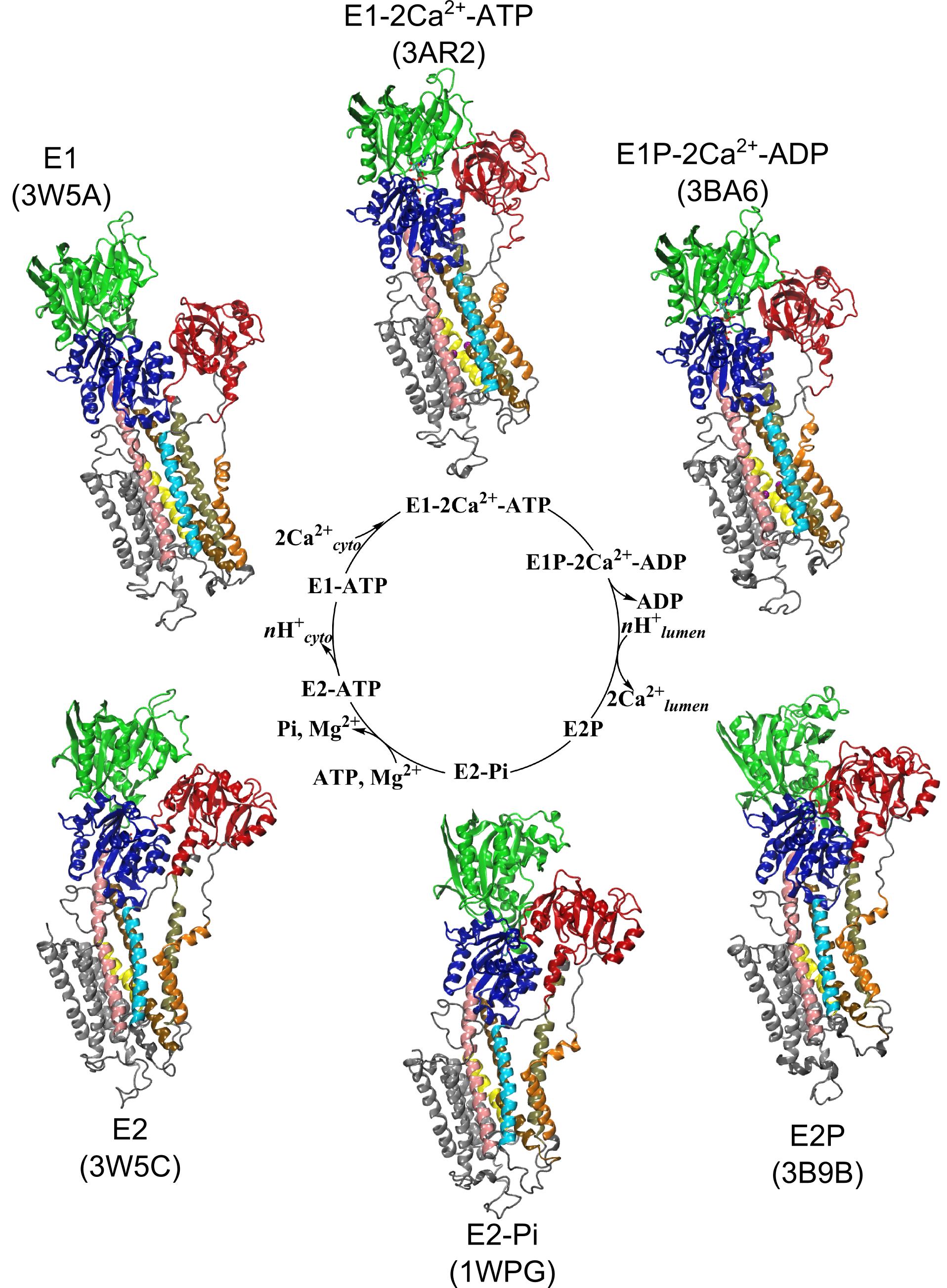
1:55 Huan Rui (University of Chicago)
Probing the cycling mechanism of the P-type ATPase: Insights from kinetic modeling simulations
2:20 Break, APS 402 Atrium

Session IV: Poster Rapid Fire

2:50 Yeong-Kyun Shin (Iowa State University)
Introduction
2:55 Poster presenters
2 minute talks

Session V: Keynote Speaker 1

4:45 A Brief Vignette Honoring Jean Chin (Eduardo Perozo, University of Chicago)
5:00 Alessio Accardi (Weill Cornell University)
Introduction
5:10 Christopher Miller (Brandeis University)
How channel crystallography made me stupid
6:00 Dinner, APS 402 – Lower Gallery
Posters, APS 402 – E1100/E1200

Saturday, November 11

Session VI: Calculating Conformational Changes: At the Bleeding Edge

7:45 Continental Breakfast, APS 402 Atrium
8:25 Wonpil Im (LeHigh University)
Introduction
8:30 Benoit Roux (University of Chicago)
C-type Inactivation and the Constricted-Like Conformations of the Selectivity Filter of K+ Channels
8:55 Michael Grabe (University of California, San Francisco)
TMEM16 lipid scrambles bend membranes to get things done
9:20 Jose Faraldo-Gomez (National Institutes of Health)
The Dos and Don’ts of the Alternating-Access Mechanism: Lessons from the Sodium-Calcium Exchanger
9:45 Break, APS 402 Atrium

Session VII: Protein Motion Within a Field: Ins and Outs of Voltage-dependent Gating

10:25 Sudha Chakrapani (Case Western Reserve University)
Introduction
10:30 Jian Yang (Columbia University)
Cryo-EM structure of a eukaryotic cyclic nucleotide-gated channel
10:55 Bonnie Wallace (Birbeck, University of London)
Structural, Function and Disease-Related Aspects of Sodium Channel Voltage-Gating
11:20 Francisco Bezanilla (University of Chicago)
Voltage sensors and membrane capacitance
11:45 Rama Ranganthan (University of Chicago)
Protein mechanics: the link between structure, function, and evolution
12:10 Lunch, APS 402 – Lower Gallery

Session VIII: Structure and Dynamics with Few Molecules (or a Lot)

1:25 Valeria Vasquez (University of Tennessee, Memphis)
Introduction
1:30 Robert Fischetti (APS, Argonne National Lab)
Serial crystallography with monochromatic and polychromatic X-ray beams, and the APS Upgrade
1:55 Yeon-Kyun Shin (Iowa State University)
Zooming in on single vehicle fusion
2:20 Simon Scheuring (Weill Cornell University)
High-Speed Atomic Force Microscopy: A New Tool for the Study of the Dynamics of Single Unlabeled Membrane Proteins
2:45 Break, APS 402 Atrium
Posters, APS 401 – E1100/E1200

Session IX: The Good, the Bad and the Ugly: Conformational Changes and the Transport/Translocation Cycle

4:30 Ming Zhou (Baylor University)
Introduction
4:35 Filipo Mancia (Columbia University)
The Molecular Mechanisms Underlying Cellular Uptake of Vitamin A
5:00 Aurelio Galli (University of Vanderbilt)
Failure to Prime the Dopamine ‘Pump’ in Autism
5:25 Alessio Accardi (Weill Cornell University)
Exploring the backdoor of CLC channels and transporters: how a glutamate gets in and out of the Cl- permeation pathway
5:50 Dinner, APS 402 – Lower Gallery
Posters, APS 401 – E1100/E1200

Sunday, November 12

Session X: Structural Dynamics Through Imaging

9:00 Tobin Sosnick (University of Chicago)
Introduction
9:05 Christine Ziegler (Universität Regensburg)
Being in the right place: Localization-dependent lipid interactions in the PC2 TRP Channel
9:30 Irina Serysheva (University of Texas Houston)
Structure of IP3R Channel: Towards Understanding Gating Mechanism
9:55 Vera Moiseenkova-Bell (University of Pennsylvania)
Molecular mechanism of the TRPV2 channel pore dynamics during ligand activation
10:20 Mingley Zhao (University of Chicago)
Molecular Mechanism of SNARE Complex Disassembly
10:55 Break, APS 402 – Atrium

Session XI: Keynote Speaker 2

11:05 Robert Nakamoto (University of Virginia)
Introduction
11:15 Olga Boudker (Weill Cornell University)
Dynamic underpinnings of transport mechanism in glutamate transporters

Closing

 *Shuttle service from the guest house to the conference center will start 15 minutes prior to the start of the meeting.

*Shuttle service from the conference center to the guest house will run for 15 minutes after the poster session (9:00pm).

Announcing Frontiers in Membrane Protein Structural Dynamics 2017

Every other year, as part of our NIH sponsored Membrane Protein Structural Dynamics Consortium we gather in Chicago for a conference to discuss the most exciting aspects at the frontiers of membrane protein structure functional and dynamics. We (and the NIH) see this as gathering of the leaders in the field and a great opportunity to interface the advances of the Consortium with the community at large.

We are happy to announce the newest version of our very successful “Frontiers in Membrane Protein Structural Dynamics” meeting. It will take place on November 10-12 at the Advanced Photon Source Conference center at Argonne National Lab. We are still finalizing the program but it is expected to have a full slate of very exciting talks. The conference will begin on the evening of Friday, November 10th and end in the afternoon on Sunday, the 12th. There will be a total of 8 symposia sessions, and two keynote talks, bookending the meeting, plus an extensive poster session accompanied by a “flash session” where poster presenters will summarize their work in 1 slide/2 min micro talks.

As before, we hope to encourage attendance of graduate students and post-doctoral fellows by having a low non-speaker participant fee and offering travel awards to students, postdocs, and new faculty.

An on-line registration site will be available soon for this meeting at: http://memprotein.org/meeting

Looking very much forward to have you in the Chicago area in November!

With our best wishes (the organizing committee),

Eduardo Perozo
Hassane Mchaourab
Robert Nakamoto
Robert Fischetti
Olga Boudker
Chris Ahern

Emad Tajkhorshid and co-PIs at UIUC recipients of NIH High-Risk, High-Reward Research

University of Illinois professor and MPSDC team member Emad Tajkhorshid, along with co-PIs Chad Rienstra (Chemistry) and James Morrissey (Biochemistry) have been awarded a Director’s Transformative Research Award from the National Institutes of Health for their highly creative approach to the study of cell membrane lipids.

Membrane proteins are abundant in eukaryotic cells and play important roles in a great many biological processes ranging from cell adhesion and recognition to energy production to signaling cascades. 

Membrane proteins make up more than half of the targets for currently approved drugs, which underscores their relevance to human disease but less is known about the membrane lipids that interact with proteins and ligands.

It is becoming increasingly clear that lipids are effector molecules that modulate and/or directly carry out essential biological functions at very different rates depending on what types of lipids are present. Some examples include blood clotting, cell recognition (in immunological response especially), ion conduction (important for neuronal function and viral infection), transport of drugs across the membrane, and pain response.

A potential long-term application is the development of more effective drugs that target biological membranes. Since about 60% of the drugs on the market target membrane-bound proteins, a better understanding of lipid structure and dynamics could greatly improve the efficacy of drug design efforts by modeling the interactions that take place. This would have broader impacts on understanding all the biological functions above and potentially to address the resulting pathologies or diseases. Better blood thinners would help to ameliorate deep vein thrombosis, heart attacks and strokes. Improved modeling of immunological cell recognition and viral life cycles would help to address infectious diseases ranging from influenza to HIV/AIDS. Understanding how drugs are transported would aid in the development of better antibiotics. The project aims to develop a toolkit of methods that would be available to researchers addressing this range of problems and many others.

The High-Risk, High-Reward Research (HRHR) program, supported by the National Institutes of Health (NIH’s Common Fund) awarded twelve transformative research awards funded by the Director’s office. The awards span the broad mission of the NIH and include groundbreaking research.

Read more about the project here: link

Conformational transitions and alternating-access mechanism in the sarcoplasmic reticulum calcium pump

Avisek Das, Huan Rui, Lei Huang, Robert Nakamoto & Benoît Roux

http://www.sciencedirect.com/science/article/pii/S0022283617300219

Ion pumps are integral membrane proteins responsible for transporting ions against concentration gradients across biological membranes. Sarco/endoplasmic reticulum Ca2+-ATPase (SERCA), a member of the P-type ATPases family, transports two calcium ions per hydrolyzed ATP molecule via an “alternating-access” mechanism. While X-ray crystallography provides high-resolution snapshots about the stable experimentally resolved states of the transport cycle (Figure 1), it is very difficult to detect the short-lived intermediate conformations occurring transiently during the transport cycle.

Figure 1: Structures of relevant functional states. The transport cycle of SERCA along with the structures of functional states used in the present work. The Protein Data Bank (PDB) id of each state is given in parenthesis below the state name.

Computational methods can be used to help supplement the missing information by providing atomic models for the transient intermediates along the transport cycle. Our goal with the present effort was to elucidate the details of the alternating access mechanism in SERCA by simulating the large-scale conformational transitions between the experimentally resolved stable sates. We calculated the pathway for three key steps:

  • Cytoplasmic calcium binding and occlusion (E1 to E1-2Ca2+-ATP). Under normal physiological conditions, the pump is activated by the binding of Ca2+ ions and ATP, leading to the occlusion of the pump and the hydrolysis of ATP. A complete chronology of the binding of Ca2+ ions and the concomitant large-scale conformational changes leading to the occluded state are provided by the string pathway between the E1 and the E1-2Ca2+-ATP states.
  • Luminal opening and calcium release (E1P-2Ca2+-ADP to E2P). Once the protein is stably locked in the occluded E1-2Ca2+-ATP state for an extended period of time, the phosphoryl transfer chemical reaction between ATP and Asp351 is expected to occur spontaneously, bringing the pump to the E1P-2Ca2+-ADP state (this reaction is not simulated here). This leads us to the next step in the transport cycle, which is the isomerization and conversion of the phosphorylated pump into the E2P state, where the luminal access channel is open.
  • Closing of luminal gate and dephosphorylation (E2P to E2-Pi). After the release of Ca2+ ions, the next step in the transport cycle corresponds to the transition from the E2P state to the E2-Pi state, comprising two important molecular events: the closing of the luminal gate, and the dephosphorylation of Asp351 in the P domain.

Summary

The computational pathways provide a clear chronology of the key events underlying the active transport of calcium ions by SERCA. The main conclusions regarding three critical steps of the transport cycle E1→E1-2Ca-ATP→E2P→E2-Pi are:

  • (E1 to E1-2Ca-ATP) Starting from the cytoplasmic open-access E1 state, the binding of ATP and the formation of the phosphoryl transfer catalytic site at the interface of the N and P domain together with the binding and sequestration of the two Ca2+ ions in the TM binding sites leading to occlusion are highly concerted and cooperative. The highly concerted transition explains the tight coupling between Ca2+ binding and ATP hydrolysis that is observed experimentally.
  • (E1-2Ca-ATP to E2P) Phosphorylation of Asp351 when the pump is in the occluded E1-2Ca-ATP state drives the opening of the luminal gate via a two-step mechanism. In a first step, phosphorylation of Asp351 triggers a large-scale rotational movement of the cytoplasmic A domain to form a strong interaction with Asp351-P at the surface of the P domain (blocking of the catalytic site and preventing the back reaction by the ADP). In a second step, the large movement of the A domain is subsequently transmitted to the M4 helix via M3 helix leading to the disruption of the Ca2+ binding sites and the opening of the TM region toward the luminal side.
  • (E2P to E2-Pi) The closing of the luminal gate following the dissociation of the Ca2+ ions to the luminal solution and protonation of binding sites residues is correlated with the dephosphorylation of Asp351-P via a two-step mechanism. In a first step, dehydration of the (protonated) binding sites together with the hydrophobic re-packing between the TM helices M4, M5 and M6 spontaneously drives the closing of the luminal gate. In a second step, the closed luminal gate mediated by the junction with the M2 helix induces a rotation in the position of the A domain, positioning the 181TGES loop in the A domain together with increased water access near Asp351-P needed for the dephosphorylation reaction.

Acknowledgments

This work was supported by the Membrane Protein Structural Dynamics Consortium funded by NIH/NIGMS through grant U54-GM087519. An award of computer time was provided by the Innovative and Novel Computational Impact on Theory and Experiment (INCITE) program from the Department of Energy (DOE) of the United States of America. This research used resources of the Argonne Leadership Computing Facility (ALCF), which is a DOE Office of Science User Facility supported under Contract DE-AC02-06CH11357. Additional computational resources were provided by the Great Lake Consortium for Petascale Computing and the XSEDE program of the National Science Foundation of the United States of America.

Third Coast Workshop on Biological Cryo-EM announced for March 3, 2017

March 3, 2017 • 9:00am – 6:00pm
Saieh Hall, University of Chicago

In the past few years, a revolution in cryo electron microscopy has taken structural biology by storm. The recent integration of new developments in electron microscopes, direct electron detection cameras, and advances in image analysis methods are allowing the expansion of high resolution structural molecular biology in new and exciting directions by direct visualization of macromolecules and their complexes. The next decades will be dominated by the study of protein-protein, protein-nucleic acid complexes, molecular machines, and their conformational changes in ways that were impossible before due to their size and/or the need to study them in crystalline form. In addition, developments in many aspects of electron microscopy are providing new tools for the study of biological molecules from the single molecule to the cellular level. The Third Coast Workshop on Biological Cryo-EM will address key developments in this fast advancing field and will provide scientists from different disciplines with an opportunity to discuss the state of the field and exchange views from both experimental and computational perspectives.

Program, registration, and poster at the website: http://memprotein.org/cryoem-workshop/


Categories
Recent Posts
Archives
Recent Comments
Recent Photos