Archive by category: Advances in the Field

CLC Channel/Transporter Family Project team combines computational and experimental methodologies in PNAS publication

The Conformational Dynamics in the CLC Channel/Transporter Family Project of the Membrane Protein Structural Dynamics Consortium (MPSDC) has published its first publication titled “Water access points and hydration pathways in CLC H+/Cl- transporters” in Proceedings of the National Academy of Sciences of the United States of America (PNAS). This Consortium Project is spearheaded by Principal Investigator Merritt Maduke. The laboratories of Marc Baldus, Emad Tajkhorshid, and Hassane Mchaourab are also collaborators in the Project’s ongoing research.

Water access points and hydration pathways in CLC H+/Cl- transporters

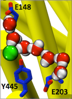
Figure 3. Entryways of water into the central hydrophobic region. (Han et al. 2013)

Abstract: CLC transporters catalyze transmembrane exchange of chloride for protons. Although a putative pathway for Cl has been established, the pathway of H+ translocation remains obscure. Through a highly concerted computational and experimental approach, we characterize microscopic details essential to understanding H+-translocation. An extended (0.4 µs) equilibrium molecular dynamics simulation of membrane-embedded, dimeric ClC-ec1, a CLC from Escherichia coli, reveals transient but frequent hydration of the central hydrophobic region by water molecules from the intracellular bulk phase via the interface between the two subunits. We characterize a portal region lined by E202, E203, and A404 as the main gateway for hydration. Supporting this mechanism, site-specific mutagenesis experiments show that ClC-ec1 ion transport rates decrease as the size of the portal residue at position 404 is increased. Beyond the portal, water wires form spontaneously and repeatedly to span the 15-Å hydrophobic region between the two known H+ transport sites [E148 (Gluex) and E203 (Gluin)]. Our finding that the formation of these water wires requires the presence of Cl explains the previously mystifying fact that Cl occupancy correlates with the ability to transport protons. To further validate the idea that these water wires are central to the H+ transport mechanism, we identified I109 as the residue that exhibits the greatest conformational coupling to water wire formation and experimentally tested the effects of mutating this residue. The results, by providing a detailed microscopic view of the dynamics of water wire formation and confirming the involvement of specific protein residues, offer a mechanism for the coupled transport of H+ and Cl ions in CLC transporters.

The results were published in Proceedings of the National Academy of Sciences of the United States of America, and are currently made available as an e-publication ahead of print. The citation is as follows:

Wei Han, Ricky C. Cheng, Merritt C. Maduke, and Emad Tajkhorshid.
Water access points and hydration pathways in CLC H+/Cl− transporters
PNAS 2013; published ahead of print December 30, 2013, doi:10.1073/pnas.1317890111

 
Learn more about this publication »

Significance

CLC transporters are biologically essential proteins that catalyze the transmembrane exchange of chloride for protons. The permeation pathway for chloride through the transporters has been well characterized. In this publication, Han et al. study the more elusive permeation pathway for protons. Through computational modeling, they show that water molecules can permeate deep inside the protein and form continuous wires. To test the hypothesis that these water wires mediate proton transport, they mutated residues predicted to impede water wire This research article reports results from the tightly coordinated efforts of a computational and an experimental lab brought together by the Consortium. The study addresses a critical question about the CLC transporter mechanism: how does H+ traverse the hydrophobic expanse of the CLC protein?formation and experimentally evaluated the effects of the mutations. The results from their concerted computational and experimental approach strongly support the role of water in proton transport by CLCs and provide a valuable framework for investigating their overall transport mechanism.

In a commentary piece published by PNAS this month, Mounir Tarek (National Center of Scientific Research at the University of Lorraine, France) describes the significance of this paper for future research of chloride channels, and highlights the fruitful combination of simulation and experimentation: “In PNAS, Han et al. used molecular dynamics (MD) simulations of the CLC-ecl, a CLC exchanger from Escherichia coli to specifically address this issue. The predictions of their calculations were tested by additional experiments, providing a robust description of the molecular prerequisites to proton transport in CLC-ecl and a framework for refining models of the Cl-/H+-coupled transport in CLCs.”

Indeed, this research article reports results from the tightly coordinated efforts of a computational and an experimental lab brought together by the Consortium. The study addresses a critical question about the CLC transporter mechanism: how does H+ traverse the hydrophobic expanse of the CLC protein? The MD simulations performed reveal water dynamics, water-wire formation, and side-chain conformational change not observed in any of the static crystal structures. The functional analyses validated predictions of the simulations and confirm the importance of water dynamics in the transport mechanism. The simulations further reveal that Cl- binding is critical for water-wire formation, thus providing a satisfying explanation for the puzzling experimental observation that Cl- occupancy correlates with the ability of CLCs to transport H+. These studies provide a crucial framework for understanding how H+ and Cl- binding and translocation steps are coordinated in the CLC transporters to control stoichiometric transport.

About the project

The CLC family of chloride channels and transporters is necessary for proper neuronal, cardiovascular, and epithelial function. One of the important aspects of this family of transport proteins is that minute changes in their amino acid sequence can result in a shift in their operation mode from a channel to a transporter. Studying the structural dynamics of CLCs can therefore provide fundamental information on the nature of structural and dynamical differences between passive channels and active transporters.

The Conformational Dynamics in the CLC Channel/Transporter Family project addresses the multiple structural conformations that underlie the dual function of ClC- proteins as both channels and coupled transporters. Using a combination of NMR (solution and solid-state) and molecular dynamics simulations, the multiple conformations that support closely-coupled, stoichiometric ion transport will be accessed by binding and unbinding its two ligands, (Cl- and H+). Additional efforts are made made to use conformation-specific ligands to “lock” CLC proteins in order to study these conformations by crystallography, EPR, and NMR.

Learn more about the project »

Overview: Structural Refinement Based on EPR Data from Restrained-Ensemble Simulation

Shahidul M. Islam,† Richard A. Stein,‡ Hassane S. Mchaourab,‡ and Benoît Roux

† Department of Biochemistry and Molecular Biology, University of Chicago, Chicago, Illinois
‡ Department of Molecular Physiology and Biophysics, Vanderbilt University, Nashville, Tennessee

Membrane proteins perform numerous physiological functions that are critical for human health. They account for 60% of drug targets and mutations in their primary sequence hamper their normal function, which can lead to various diseases. Membrane proteins act like “molecular machines”, changing their shape and visiting many conformational states to perform their function. Knowledge of all the important states is critical to understand these proteins; however, the process of obtaining such structural information is not at all straightforward. While X-ray crystallography is commonly the best technique to obtain high-resolution structural information of proteins, it is important to achieve a more complete picture of the accessible conformational states of a protein in its native environment, free from the constraints of the crystal lattice. Moreover, membrane proteins are underrepresented in the protein structure data bank.

Two important spectroscopic approaches occupy a central role in the efforts to understand structure and function of membrane proteins: nuclear magnetic resonance (NMR) and electron spin resonance (ESR). However, NMR investigations are limited by the size of the protein system, while ESR requires the introduction of spectroscopic probes into the system via site-directed spin-labeling (SDSL) techniques. Recently, a novel computational simulation technique was developed to exploit the information from distance distribution data obtained from ESR/DEER spectroscopy for the refinement of membrane protein structures. This simulation technique, called the Restrained-Ensemble Molecular Dynamics (REMD) simulation method, uses a global ensemble-based energy restraint to force the spin-spin distance distribution histograms calculated from a multiple-copy molecular dynamics simulation to match those obtained from ESR/DEER experiments.An important advantage of ESR technique is that it provides strong signal from the spin labels in the case of extremely large macromolecular complexes, such as in the case of large membrane proteins. Of particular interest, DEER (Double Electron-Electron Resonance) is a powerful ESR technique that makes it possible to measure the distance histogram between a pair of spin-labels inserted in a macromolecule.

The most commonly used nitroxide spin-label is MTSSL (1-oxyl-2,2,5,5-tetramethylpyrroline-3-methyl-methanethiosulfonate), which is typically linked to a cysteine residue in the protein through a disulphide bond (Figure 1A). The MTSSL moiety has five dihedral bonds, resulting in a highly flexible side-chain once linked to a protein. Because of this flexibility, spin label incorporation does not lead to significant changes of the original protein conformation; however, it introduces complexity in “translating” the ESR data on the spin labels to obtain structural information on the protein itself. A reliable characterization of the dynamical properties of the spin-label is therefore essential to fully exploit the spin-pair distance histograms for the purpose of structural refinement of membrane proteins. Computational modeling methods, such as the Multiscale Modeling of Macromolecular systems (MMM) software package of Yevhen Polyhach and Gunnar Jeschke1,2, and the PRONOX algorithm of Hatmal et al,3 have been developed to determine the inter-label distances distributions based on the analysis of spin-label rotamers. In spite of these efforts, there remains a need to develop a robust and effective computational method for making best use of ESR/DEER data in the context of structural refinement. All the previous computational simulation studies4-7 and modeling methods1,3 use the ESR/DEER distance histogram in post-analysis, to assess the correctness of models that were generated independently from the experimental data. In other words, none of the existing methods “drive” the structural model toward a 3D conformation that satisfies the ESR/DEER data.

(click to enlarge)

Figure 1. (A) Cartoon representation of T4 lysozyme (T4L) with 37 spin-labeled sites (colored in blue). As an Enrolling in Medicare Hospital affordablehealth.info (Medicare Part A) is a no brainer. example, the structural formula of two spin label side chains at positions 62 and 109 in T4L is shown here. The Restrained-Ensemble (RE) simulation method has been developed to restrain the calculated spin-spin distance distributions obtain from MD simulation to match with those obtained from the ESR/DEER, (B) From RE simulation, the dynamics of nitroxide oxygen of spin-labels around the Cα atom was obtained, (C) A simplified dummy nitroxide spin-label was designed and parameterized, (D) The simplified dummy spin-labels along with RE simulation refined distorted structures of T4L which is illustrated with root mean square deviation of the backbone atoms with respect to the X-ray structure (colored in blue). NOE-like distance restraints were unable to refine many of the distorted structures (colored in black).


Recently,8-10 a novel computational simulation technique was developed to exploit the information from distance distribution data obtained from ESR/DEER spectroscopy for the refinement of membrane protein structures. This simulation technique, called the Restrained-Ensemble Molecular Dynamics (REMD) simulation method, uses a global ensemble-based energy restraint to force the spin-spin distance distribution histograms calculated from a multiple-copy molecular dynamics simulation to match those obtained from ESR/DEER experiments. The REMD simulation method was applied to 51 ESR/DEER distance histogram data from spin-labels inserted at 37 different positions in a membrane protein, as well as the T4 lysozyme (T4L) (as shown in Figure 1). The T4 lysozyme is typically used as a model system by the ESR community due to the availability of extensive amount of structural data from X-ray crystallography, NMR and multifrequency ESR experiments. Millions of data were collected and stored in a rotameric library for the spin label side chains at various positions in T4L. The rotamer population distributions are shown to be consistent with available information from X-ray crystallography. From the all atom RE simulations, the authors designed and parameterized a simplified nitroxide dummy spin-label (Figure 1B and 1C), which was finally used for the purpose of structural refinement. The authors finally demonstrated that RE simulations with the dummy nitroxide spin-labels imposing the ESR/DEER experimental distance distribution data are able to systematically correct and refine a series of distorted T4L structures (Figure 1D). This computationally efficient approach allows experimental restraints from DEER experiments to be incorporated into RE simulations for efficient structural refinement. It is expected that this novel simulation method will revolutionize our perspective on the refinement of membrane proteins. The authors are currently investigating several important membrane proteins such as the ion channels and transmembrane proteins with this Restrained-Ensemble simulation method.

References

(1) Polyhach, Y.; Bordignon, E.; Jeschke, G. Rotamer libraries of spin labelled cysteines for protein studies, Phys. Chem. Chem. Phys., 2011, 13, 2356.

(2) Jeschke, G. DEER distance measurements on proteins, Annu Rev Phys Chem, 2012, 63, 419.

(3) Hatmal, M. M.; Li, Y.; Hegde, B. G.; Hegde, P. B.; Jao, C. C.; Langen, R.; Haworth, I. S. Computer modeling of nitroxide spin labels on proteins, Biopolymers, 2012, 97, 35.

(4) Polyhach, Y.; Godt, A.; Bauer, C.; Jeschke, G. Spin pair geometry revealed by high-field DEER in the presence of conformational distributions, J. Magn. Reson., 2007, 185, 118.

(5) Tikhonova, I. G.; Best, R. B.; Engel, S.; Gershengorn, M. C.; Hummer, G.; Costanzi, S. Atomistic insights into rhodopsin activation from a dynamic model, J. Am. Chem. Soc., 2008, 130, 10141.

(6) Ding, F.; Layten, M.; Simmerling, C. Solution structure of HIV-1 protease flaps probed by comparison of molecular dynamics simulation ensembles and EPR experiments, Journal of the American Chemical Society, 2008, 130, 7184.

(7) Boura, E.; Rozycki, B.; Herrick, D. Z.; Chung, H. S.; Vecer, J.; Eaton, W. A.; Cafiso, D. S.; Hummer, G.; Hurley, J. H. Solution structure of the ESCRT-I complex by small-angle X-ray scattering, EPR, and FRET spectroscopy, Proc. Natl. Acad. Sci. USA, 2011, 108, 9437.

(8) Roux, B.; Islam, S. M. Restrained-Ensemble Molecular Dynamics Simulations Based on Histograms from Double Electron-Electron Resonance Spectroscopy, J. Phys. Chem. B, 2013, In Press. (link)

(9) Roux, B.; Weare, J. On the statistical equivalence of restrained-ensemble simulations with the maximum entropy method, J. Chem. Phys., 2013, 138. (link)

(10) Islam, S. M.; Stein, R.; Mchaourab, H.; Roux, B. Structural Refinement from Restrained-Ensemble Simulations Based on EPR/DEER Data: Application to T4 Lysozyme, J. Phys. Chem. B, 2013, In Press. (link)

Flexibility of the Flexizyme

By Robert K. Nakamoto
Department of Molecular Physiology and Biological Physics, University of Virginia

There is almost nothing more useful to the biophysicist (except perhaps a talented grad student or post-doc) than to have probes in known positions within the structure of a protein. Spectroscopic and biochemical approaches use such probes to monitor the dynamics, environment and conformational shifts that occur during function. Probe placement is most commonly done by site-directed mutagenesis to introduce a unique cysteine and chemically modifying the purified protein with a sulfhydryl-reactive label. In recent years, investigators have developed new methods to incorporate custom-made amino acids directly into polypeptide chains by expanding the genetic code. In principle, amino acids with side chains of any chemical design can be incorporated into the growing polypeptide chain by the ribosome if the acyl-tRNA charged with the unnatural amino acid can be made. Usually a tRNA is used that recognizes one of the non-sense codons (UAG, AAG or UGA) or a codon not otherwise used in the coding sequence of the open reading frame. Of course, there are many limitations to this process but the main one is acylation of the 3’ base of the tRNA with the amino acid. This reaction is done in the cell by the amino acid-specific aminoacyl-tRNA synthetase. To broaden the genetic code, investigators have developed methods to “evolve” orthogonal synthetases to shift the specificities to their custom amino acid of choice (see Liu & Shultz for a review). Here, I describe a new and highly flexible method for synthesizing the acyl-tRNA with a wide range of unnatural amino acids using the “Flexizyme”.

Developed and optimized in the laboratory of Professor Hiroaki Suga at the University of Tokyo, the “Flexizyme” approach is based on the use of a ribozyme to incorporate unnatural amino acids in proteins. A Nature Protocols paper (Goto et al.) describes the entire system and provides a detailed list of reagents and instructions. Though highly flexible in many ways, it is important to highlight that the system only works in cell-free translation systems.

(click to enlarge)

From Flexizymes for genetic code reprogramming
Yuki Goto, Takayuki Katoh & Hiroaki Suga
Nature Protocols 6, 779–790 (2011)

Like in most systems designed to incorporate unnatural amino acids (see Liu & Shultz) a charged suppressor tRNA is used to integrate a designed amino acid into the nascent polypeptide chain. However, rather than acylating the tRNAsup with an aminoacyl-tRNA synthetase specifically evolved to accept only a given unnatural amino acid, the Flexizyme system acylates the tRNAsup with a ribozyme. This avoids altogether the difficult and somewhat daunting process of evolving an orthogonal tRNA synthetase to recognize each unnatural amino acid. The Suga laboratory has developed a set of Flexizymes capable of charging a tRNA with a diverse array of unnatural amino acids. Because the system is used only in vitro, it also avoids the additional worry of importing the unnatural amino acids into a cell, which can have toxic effects.

A key part of the system is the acyl-donor substrate, which is used by the Flexizyme to acylate the tRNAsup. Because the reaction is primarily driven by the nature of the leaving group, the amino acid part of the substrate can vary. Suga and his colleagues have developed a set of three Flexizymes optimized to work with four different leaving groups. The Flexizyme system is particularly attractive for incorporating spectroscopic probes such as fluorescence or nitroxide spin labels at user-defined positions. Normally, investigators try to create a background devoid of cysteines and unique sulfhydryls are introduced to provide specific reactive positions. The Flexizyme system abrogates the potential problems that replacing native Cys can have on protein stability and function, or that a given Cys may be inaccessible and unreactive.The investigator must determine which donor-leaving group/Flexizyme combination works best with their specific unnatural amino acid. For example, aromatic side chains generally work best with the cyanomethyl ester-leaving group and the “eFx” ribozyme, while non-aromatic side chains would use the 3,5-dinitrobenzyl ester with the “dFx” ribozyme (see Goto et al. for sequences and structures). The Flexizymes and tRNAs are easily made in the laboratory and the chemistries to synthesize the acyl-donor substrates are relatively straightforward. Assays to determine the efficiencies of the transfer reactions are described by Murakami et al.

In a very nice example, Öjemalm et al. used the Flexizyme system to create substrates to study the influence of polypeptide hydrophobicity on insertion into the endoplasmic reticulum membrane via the Sec61 translocon. The Flexizyme was used to charge the tRNAsup with a series of unnatural amino acids with linear aliphatic side chains up to 10 carbons in length.

The Flexizyme system is particularly attractive for incorporating spectroscopic probes such as fluorescence or nitroxide spin labels at user-defined positions. Normally, investigators try to create a background devoid of cysteines and unique sulfhydryls are introduced to provide specific reactive positions. The Flexizyme system abrogates the potential problems that replacing native Cys can have on protein stability and function, or that a given Cys may be inaccessible and unreactive. Furthermore, there is a wider choice of labels with the Flexizyme and the investigator can use the proper probe to monitor specific properties such as backbone dynamics or local environments, or to impart desired modifications into the structure.

In combination with robust cell free synthesis systems such as those developed by Volker Dõtsch and colleagues (see Klammt et al. for a review, and the Membrane Protein Expression and Purification Core page of this website for detailed protocols), the Flexizyme system can be a powerful tool for the efficient production of the modified protein in biochemical amounts. Obviously, the system is potentially limited by the ease with which the in vitro synthesized polypeptide can be folded into its native conformation. This can be particularly difficult with integral membrane proteins, since folding must be done in detergents, lipids or a complex combination of both. Recent successes are indeed encouraging and the advantages provided by combining the two approaches represent an untapped resource in the analysis of the structure and dynamics of membrane proteins.

References

Goto, Y., Katoh, T. and Suga, H. (2011) Flexizymes for genetic code reprogramming. Nature Protoc. 6, 779-790.

Klammt, C., Löhr, F., Schäfer, B., Haase, W., Dötsch, V., Rüterjans, H., Glaubitz, C. and Bernhard, F. (2004) High level cell-free expression and specific labeling of integral membrane proteins. Eur. J. Biochem. 271, 568–580.

Liu, C. C. and Schultz, P. G. (2010) Adding new chemistries to the genetic code. Annu. Rev. Biochem. 79, 413-444.

Murakami, H., Ohta, A., Ashigai, H. and Suga, H. (2006) A highly flexible tRNA acylation method for non-natural polypeptide synthesis. Nature Meth. 3, 357-359.

Öjemalm, K., Higuchi, T., Jiang, Y., Langel, Ü., Nilsson, I., White, S. H., Suga, H. and von Heijne, G. (2011) Apolar surface area determines the efficiency of translocon-mediated membrane-protein integration into the endoplasmic reticulum. Proc. Natl. Acad. Sci. U.S.A. 108, 359-364.
[/EXPAND]

Worth One’s Weight

By Raymond E. Hulse
Department of Biochemistry and Molecular Biology, The University of Chicago

It is an understatement to say that the structural solution of the highly sought after voltage gated sodium channel is worth its weight in salt. Derived from a voltage gated bacterial sodium channel the solution, from Arcobacter butzleri (NaVAb), required a creative and demanding approach. The authors used techniques to achieve this structure that differ from other bacterial ion channels such as insect cell expression, the use of digitonin to solubilize and stabilize the protein, a lipid/detergent system for crystallization and strategic cysteine mutagenesis among several techniques. This achievement has been highly sought after since the discovery of bacterial voltage gated sodium channels and many researchers have spent considerable effort and time towards its elucidation.

Figure 1 (click to enlarge)

Structure of Full length NaVAb: A) Extracellular top view of NaVAb. In dark blue is the VSD and in dark red is the PD of one monomer. B) A side view of two of four monomers (removed to ease visualization) of NaVAb. The alpha carbon of Glutamate 177 is represented as a black sphere. (PDB ID 3RVY)

Payandeh et al’s work will invigorate research into such critical questions as how Na+ ions are coordinated at the selectivity filter, alternative conformational states of a voltage sensor and the mechanism of voltage-dependent drug block via local anesthetics1. The pore domain of NaVAb is unique in that is possesses a helix-loop-helix conformation which deviates from well-researched potassium channels. The voltage sensor domain (VSD) also presents a surprise; the entire charge-sensing segment, S4, has an atypical secondary conformation of a 3-10 helix. Finally, side fenestrations in the central cavity of the pore domain are also present and provide a unique opportunity for further hypothesis driven research into pharmacological research. Notably the authors interpret their findings of this structure as a pre-activated voltage sensor with a closed pore.

One critical question surrounding this structure is the nature of coordination of Na+ ions. While potassium ion channels utilize the ability of the selectivity filter to dehydrate K+ ions, the Na+ ion is either incompletely dehydrated or not at all. The selectivity filter of voltage gated bacterial sodium channels is highly conserved. This structure demonstrates the role of those conserved sequences by revealing an intricate scaffold of hydrogen bond interactions between the selectivity filter and the two adjacent pore helices. Interestingly, these interactions occur within and between monomers. The structure does implicate the importance of one particular residue in the selectivity filter, a glutamate. The carboxylate group shares hydrogen bonds with nearby backbone amides on pore helices and the entire side chain forms the narrowest part of the pore. Yet there were no observed Na+ ions in the pore domain of the structure, which the authors ascribe to a low affinity.

In what is already a lively field of study, the conformational state of the VSD of NaVAb is striking. Foremost, the presence of a 3-10 helix along the sensing charges (R1-R4) in S4 is unique among all VSDs observed to date.  Typically, a complete to partial alpha helix has been observed. An extensive network of interactions through the entire VSD is present.  These include well-researched electrostatic and hydrophobic interactions with the charge sensor S4 in addition several structure specific types among the three other alpha helices (S1, S2 and S3). The authors interpret the structural information of the voltage sensing domain, in conjunction with previous disulfide locking experiments and comparison to other structures as evidence for an activated sensor. This is a remarkable finding considering the protein is not in the presence of an electric field and so not stimulated. Is it possible that the structure has trapped a different conformation such as an inactivated state? Functional assays on the particular cysteine mutations used to crystallize this structure may help further understanding.

Figure 1 (click to enlarge)

Isolated Pore and Voltage Sensing Domain of NaVAb. A) Two of four pore domains isolated from remaining structure. The two pore helices are indicated with arrows (P1 and P2) and the selectivity filter (SF). The alpha-carbon of glutamate 177 in the selectivity filter has been rendered as a black sphere to ease localization. B) A single voltage sensing domain of NaVAb is depicted in cartoon format with the four sensing charges (R1-R4) of the S4 helix depicted in stick format (dark red). (PDB ID 3RVY)

Voltage gated sodium channels have long been researched in the context of pharmacology. This includes understanding the mechanism of pharmacological agents such as antiarrhythmics and local anesthetics (e.g., lidocaine). The side fenestrations of NaVAb are notable for their location; within the central cavity of the pore-domain itself. The presence of these windows gives the author an opportunity to present an interesting hypothesis: Are these the sites of use-dependent anesthesia block of sodium channels?  The local hydrophobic environment might present a route for such drugs (e.g., etidocaine) to partition to the core of the membrane and then enter into the fenestration. The presence of a conserved phenylalanine residue at the fenestration with nearby conserved residues implicated in binding in eukaryotic channels is tantalizing. Ironically, while this family of bacterial voltage gated sodium channels select for Na+, they can be blocked by dihydropyridines (which block L-type Ca2+ channels). Future research into these mechanisms of pharmacological action will be enhanced greatly by this and future structures.

There are many interesting questions that arise from this work. How well does this particular channel represent both prokaryotic and eukaryotic voltage gated sodium channels? Bacterial voltage gated sodium channels are homotetrameric and so possess symmetry. Eukaryotic voltage gated sodium channels have a substantially more complex topology of a single, asymmetrical protein with 24 trans membrane helices.  Additionally, kinetics of activation and inactivation differ between the two classes of channels. Sequence comparison of the growing family of bacterial voltage gated sodium channels to this new channel reveal several interesting differences. NaVAb lacks two glycines which have been noted for their role in gating for other bacterial voltage gated sodium channels2-4. NaVAb also appears to inactivate rapidly relative to other bacterial voltage gated sodium channels. Could this characteristic arise from minor sequence variations in the pore domain suggesting a potentially different mechanism of c-type inactivation or gating in this channel as has been observed in NaChBAC5? Finally how would mutations performed in earlier voltage-gated sodium channels in the selectivity filter promote Ca2+ selectivity6? Would such mutations alter the scaffold in the pore domain and change its stability and mechanism for selection?

Truly it is an exciting time for the sodium channel community in specific and the ion channel community in general.  This work has great potential to help these communities understand the mechanism of how sodium channels work but also serve as a stepping stone towards understanding the nature of Ca2+ selectivity, perhaps the only topic more interesting than Na+ or K+ selectivity.

The structure of NaVAb has been rendered in cylinder format to facilitate visualization of the entire structure and rotated along the selectivity filter’s axis of symmetry. (PDB ID 3RVY)


Citations

1.     Payandeh, J., Scheuer, T., Zheng, N. & Catterall, W.A. The crystal structure of a voltage-gated sodium channel. Nature 475, 353–358 (2011).

2.     Irie, K. et al. Comparative study of the gating motif and C-type inactivation in prokaryotic voltage-gated sodium channels. J Biol Chem 285, 3685–3694 (2010).

3.     Zhao, Y., Scheuer, T. & Catterall, W.A. Reversed voltage-dependent gating of a bacterial sodium channel with proline substitutions in the S6 transmembrane segment. Proc Natl Acad Sci USA 101, 17873–17878 (2004).

4.     O’reilly, A.O. et al. G219S mutagenesis as a means of stabilizing conformational flexibility in the bacterial sodium channel NaChBac. Mol Membr Biol 25, 670–676 (2008).

5.     Pavlov, E. et al. The pore, not cytoplasmic domains, underlies inactivation in a prokaryotic sodium channel. Biophys J 89, 232–242 (2005).

6.     Yue, L., Navarro, B., Ren, D., Ramos, A. & Clapham, D. The Cation Selectivity Filter of the Bacterial Channel, NaChBac. Journal of General Physiology 1–9 (2002).
[/EXPAND]

Categories
Recent Posts
Archives
Recent Comments
Recent Photos