This article was originally published at Genomeweb BioInform (link).
A new consortium launched with the aim of understanding the structure and dynamic function of membrane proteins will likely result in an improved informatics toolbox for modeling proteins and other biomolecules.
Last month, the National Institute of General Medical Science launched the Membrane Protein Structural Dynamics Consortium, an interdisciplinary team of researchers that aim to use biophysical and computational methods to understand the structure, function, and dynamics of membrane proteins.
The consortium, funded by a five-year $22.5 million “glue grant” from NIGMS, comprises 30 scientists from 14 institutions in four countries and is hailed as one of the largest and most comprehensive projects focused on membrane proteins to date.
Led by researchers at the University of Chicago, the consortium also includes scientists from Cornell University, Columbia University, Germany’s Johann Wolfgang Goethe-Universität, the National Institutes of Health, Stanford University, University of California-Los Angeles, University of Illinois, University of Pittsburgh, University of Toronto, University of Virginia, University of Wisconsin, the Netherlands’ Utrecht University, and Vanderbilt University.
The project is expected to help scientists better understand a wide range of ailments caused by faulty membrane proteins, such as some forms of heart disease, diabetes, and neurological and hormonal disorders. Ultimately, since more than half of drugs currently on the market target membrane proteins, the project could pave the way for developing new or improved drugs and therapies.
The project will involve a combination of structural biology, magnetic resonance, fluorescence spectroscopy, biochemistry, and biophysics techniques, and will also have a very large computational component. Rather than assign the task of computational tool development to individual labs, the consortium has opted to delegate the task to a select group of researchers, dubbed the computational core, who have considerable expertise in that arena.
Benoit Roux, a professor of biochemistry and molecular biophysics at the University of Chicago and one of the members of the core, told BioInform that the group aims to develop tools centered around four specific aims: to generate different kinds of force fields for proteins; integrate data at different scales and resolutions; identify pathways between different conformations of proteins; and interpret spectroscopic measurements, which are often “ambiguous.”
Along similar lines, Harel Weinstein, a professor of physiology and biophysics at Cornell University, said the group will develop tools for “large-scale simulations” of protein structures that realistically represent the protein and its fluid environment, as well as tools to let researchers look at protein dynamics based on known structures and to interpret “a new generation of data” from things like single molecule experiments. The team also hopes to improve on “sophisticated” tools that are currently used in computational biology labs.
“That is quite complicated and requires several levels of new thinking … in terms of the use of computational resources [such as] parallelization and new types of processors,” he told BioInform. “We are trying to develop analysis tools that reduce the data to the same kinds of components [that] everybody [can] understand — even those who are not using the [same] experimental tool[s] for data acquisition.”
Roux said that the developers plan to use existing tools that they have developed in their labs, which they will “extend” to work in a more “general fashion.”
“It’s a challenge because [the proteins are] a bit more disparate … you have a channel, a pump, you have maybe a receptor … being analyzed,” he said. “The tool has to be general enough that it’s suitable for all these problems but not so general that it becomes a bit abstract.”
An additional challenge for the core, Weinstein said, will be to develop tools that can be easily used by experimental biologists involved in the consortium who don’t have intensive bioinformatics training. On that score, he said the consortium is considering conducting workshops that will provide training on how to use the tools.
The computational core also plans to develop a centralized database to store and integrate project data in a format that consortium members can make sense of no matter what tool was used to collect the experimental data, Weinstein said.
The planned database will be different from repositories like the Protein Structure Initiative’s Structural Biology Knowledgebase in the sense that “the same people who create the tools for dissemination and disseminate the data are the people who collect the data” he said.
“I could put my data in [PSI]; it’s a very useful tool,” Weinstein acquiesced. “What is different is [the consortium's database] is created for people who work together and who refine the database all the time as they work.”
The PSI knowledgebase is a product of the Protein Structure Initiative launched by NIGMS in 1999 with the long-term goal of making it easy to obtain three-dimensional atomic-level structures of proteins by looking at their DNA sequences.
Filling the Gap
Computational core member Ivet Bahar, a professor of computational biology at the University of Pittsburgh, told BioInform that her team plans to use “elastic network models” to analyze membrane proteins without compromising the resolution.
She said that although her models, developed over a decade ago, have been used to study other molecules, reworking these tools to work for membrane proteins is a greater challenge because they have to take into account the membrane as well as how the protein interacts with the membrane, among other things.
Membrane proteins, along with the lipid and water molecules that surround and interact with them, create large complex systems that are difficult to model, explained Bahar.
“My lab simplifies the analysis of such complex systems by using simplified models … [that] help us understand the mechanisms [and] the machinery of [the] biological processes,” she said.
According to Bahar, the models her team develops “approximate” proteins and other biomolecules as “elastic materials” such as beads and elastic springs, which provides insights into which structures are the most “energetically favorable” for different functions.
Read more at Genomeweb Bioinform »
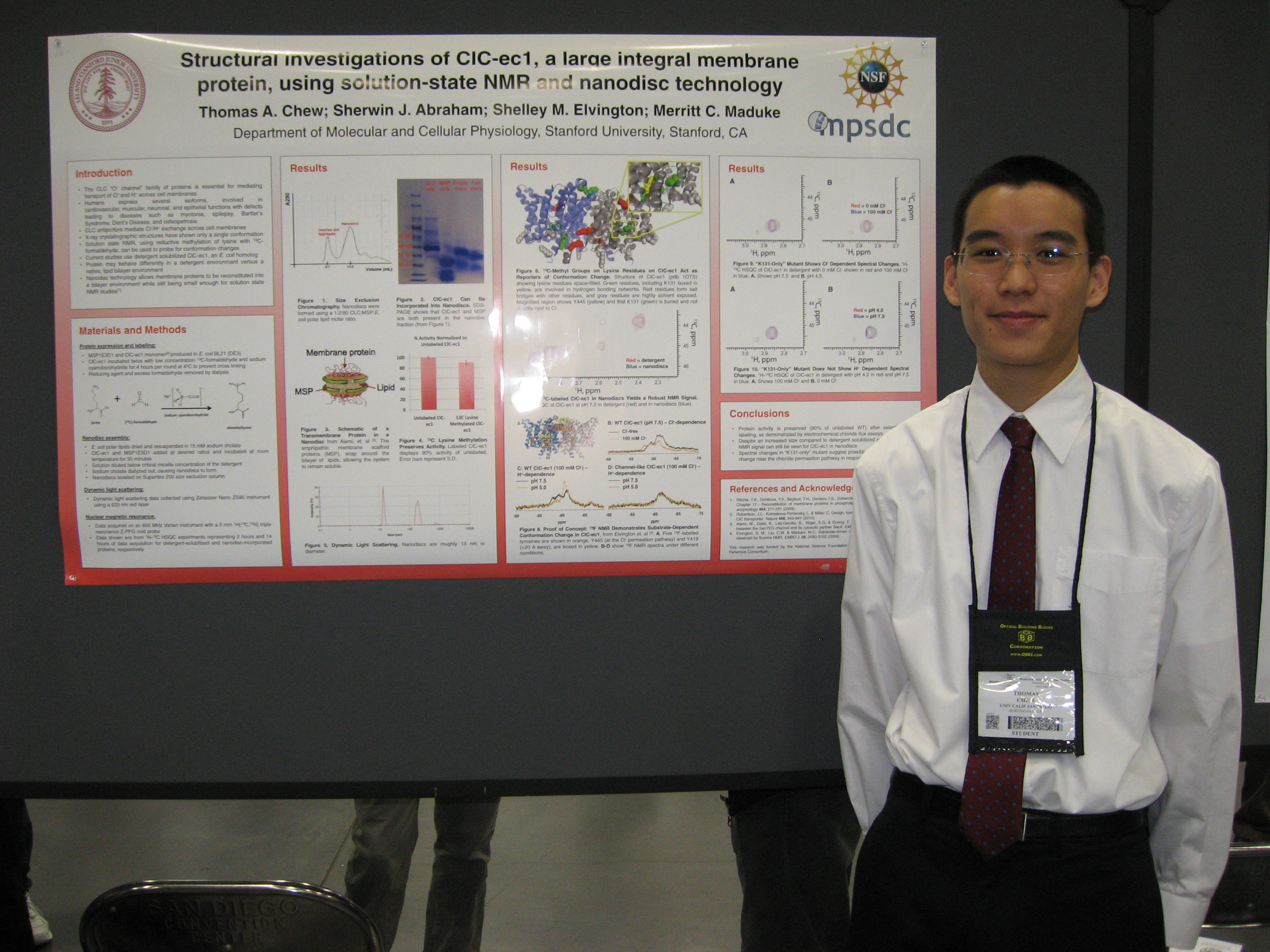 Thomas Chew with the award-winning poster. Click to enlarge.
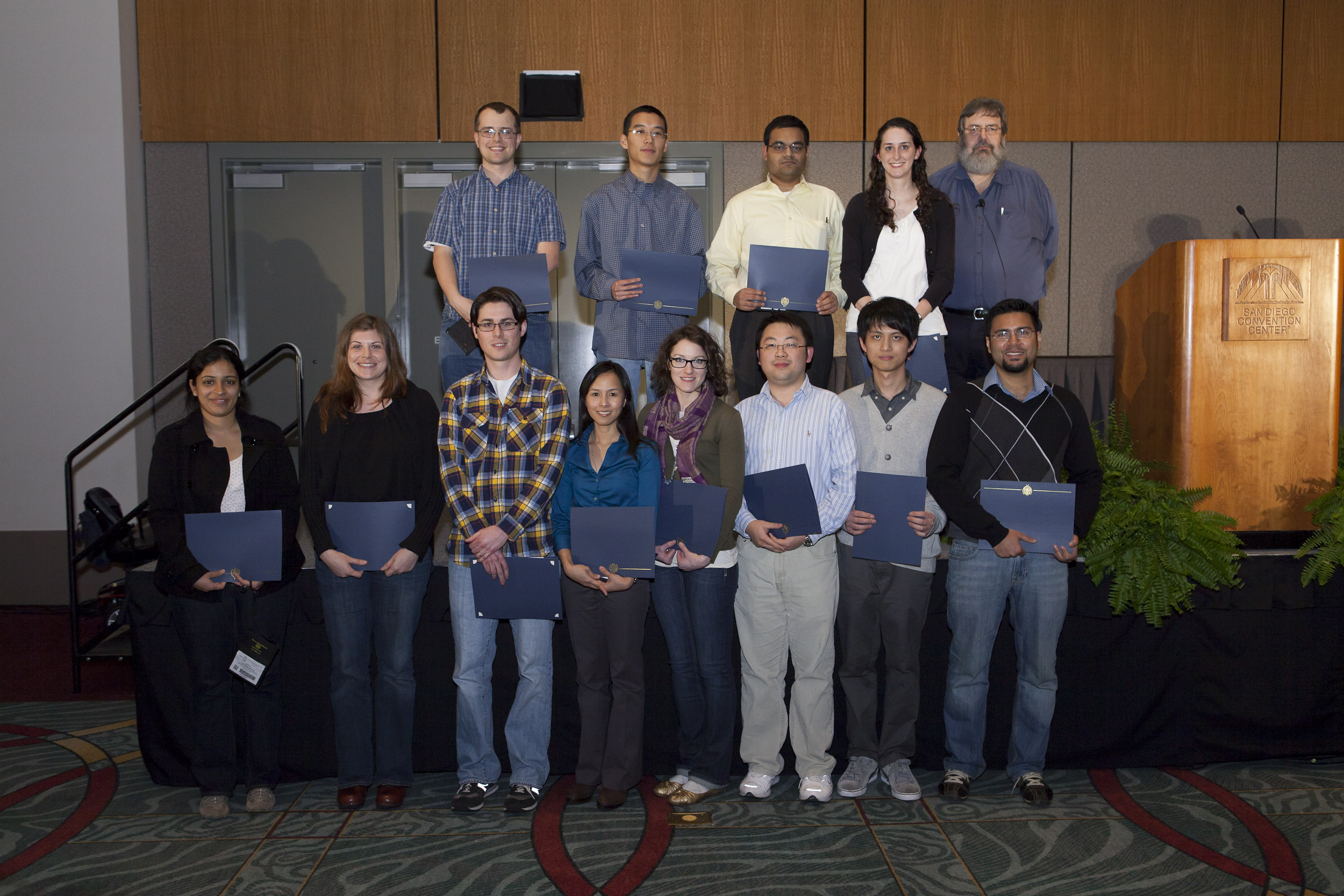 Thomas Chew at the February 27 Awards Ceremony together with the other winners and Steve Block. Click to enlarge.
 Dr. Klaus Schulten


