Emad Tajkhorshid and co-PIs at UIUC recipients of NIH High-Risk, High-Reward Research

University of Illinois professor and MPSDC team member Emad Tajkhorshid, along with co-PIs Chad Rienstra (Chemistry) and James Morrissey (Biochemistry) have been awarded a Director’s Transformative Research Award from the National Institutes of Health for their highly creative approach to the study of cell membrane lipids.

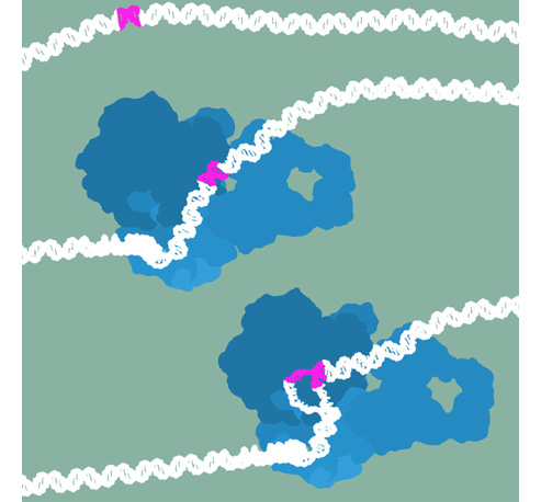
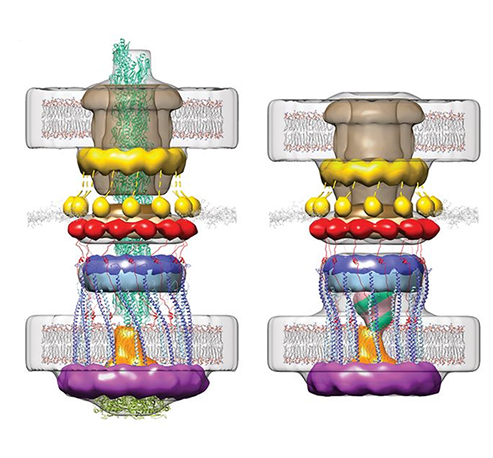
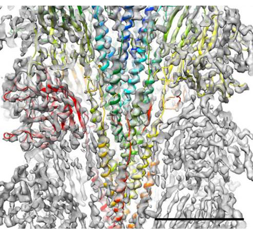
Membrane proteins are abundant in eukaryotic cells and play important roles in a great many biological processes ranging from cell adhesion and recognition to energy production to signaling cascades. 

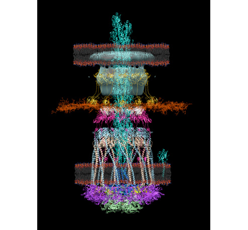
Membrane proteins make up more than half of the targets for currently approved drugs, which underscores their relevance to human disease but less is known about the membrane lipids that interact with proteins and ligands.

It is becoming increasingly clear that lipids are effector molecules that modulate and/or directly carry out essential biological functions at very different rates depending on what types of lipids are present. Some examples include blood clotting, cell recognition (in immunological response especially), ion conduction (important for neuronal function and viral infection), transport of drugs across the membrane, and pain response.

A potential long-term application is the development of more effective drugs that target biological membranes. Since about 60% of the drugs on the market target membrane-bound proteins, a better understanding of lipid structure and dynamics could greatly improve the efficacy of drug design efforts by modeling the interactions that take place. This would have broader impacts on understanding all the biological functions above and potentially to address the resulting pathologies or diseases. Better blood thinners would help to ameliorate deep vein thrombosis, heart attacks and strokes. Improved modeling of immunological cell recognition and viral life cycles would help to address infectious diseases ranging from influenza to HIV/AIDS. Understanding how drugs are transported would aid in the development of better antibiotics. The project aims to develop a toolkit of methods that would be available to researchers addressing this range of problems and many others.

The High-Risk, High-Reward Research (HRHR) program, supported by the National Institutes of Health (NIH’s Common Fund) awarded twelve transformative research awards funded by the Director’s office. The awards span the broad mission of the NIH and include groundbreaking research.

Read more about the project here: link

Conformational transitions and alternating-access mechanism in the sarcoplasmic reticulum calcium pump

Avisek Das, Huan Rui, Lei Huang, Robert Nakamoto & Benoît Roux

http://www.sciencedirect.com/science/article/pii/S0022283617300219

Ion pumps are integral membrane proteins responsible for transporting ions against concentration gradients across biological membranes. Sarco/endoplasmic reticulum Ca2+-ATPase (SERCA), a member of the P-type ATPases family, transports two calcium ions per hydrolyzed ATP molecule via an “alternating-access” mechanism. While X-ray crystallography provides high-resolution snapshots about the stable experimentally resolved states of the transport cycle (Figure 1), it is very difficult to detect the short-lived intermediate conformations occurring transiently during the transport cycle.

Figure 1: Structures of relevant functional states. The transport cycle of SERCA along with the structures of functional states used in the present work. The Protein Data Bank (PDB) id of each state is given in parenthesis below the state name.

Computational methods can be used to help supplement the missing information by providing atomic models for the transient intermediates along the transport cycle. Our goal with the present effort was to elucidate the details of the alternating access mechanism in SERCA by simulating the large-scale conformational transitions between the experimentally resolved stable sates. We calculated the pathway for three key steps:

  • Cytoplasmic calcium binding and occlusion (E1 to E1-2Ca2+-ATP). Under normal physiological conditions, the pump is activated by the binding of Ca2+ ions and ATP, leading to the occlusion of the pump and the hydrolysis of ATP. A complete chronology of the binding of Ca2+ ions and the concomitant large-scale conformational changes leading to the occluded state are provided by the string pathway between the E1 and the E1-2Ca2+-ATP states.
  • Luminal opening and calcium release (E1P-2Ca2+-ADP to E2P). Once the protein is stably locked in the occluded E1-2Ca2+-ATP state for an extended period of time, the phosphoryl transfer chemical reaction between ATP and Asp351 is expected to occur spontaneously, bringing the pump to the E1P-2Ca2+-ADP state (this reaction is not simulated here). This leads us to the next step in the transport cycle, which is the isomerization and conversion of the phosphorylated pump into the E2P state, where the luminal access channel is open.
  • Closing of luminal gate and dephosphorylation (E2P to E2-Pi). After the release of Ca2+ ions, the next step in the transport cycle corresponds to the transition from the E2P state to the E2-Pi state, comprising two important molecular events: the closing of the luminal gate, and the dephosphorylation of Asp351 in the P domain.

Summary

The computational pathways provide a clear chronology of the key events underlying the active transport of calcium ions by SERCA. The main conclusions regarding three critical steps of the transport cycle E1→E1-2Ca-ATP→E2P→E2-Pi are:

  • (E1 to E1-2Ca-ATP) Starting from the cytoplasmic open-access E1 state, the binding of ATP and the formation of the phosphoryl transfer catalytic site at the interface of the N and P domain together with the binding and sequestration of the two Ca2+ ions in the TM binding sites leading to occlusion are highly concerted and cooperative. The highly concerted transition explains the tight coupling between Ca2+ binding and ATP hydrolysis that is observed experimentally.
  • (E1-2Ca-ATP to E2P) Phosphorylation of Asp351 when the pump is in the occluded E1-2Ca-ATP state drives the opening of the luminal gate via a two-step mechanism. In a first step, phosphorylation of Asp351 triggers a large-scale rotational movement of the cytoplasmic A domain to form a strong interaction with Asp351-P at the surface of the P domain (blocking of the catalytic site and preventing the back reaction by the ADP). In a second step, the large movement of the A domain is subsequently transmitted to the M4 helix via M3 helix leading to the disruption of the Ca2+ binding sites and the opening of the TM region toward the luminal side.
  • (E2P to E2-Pi) The closing of the luminal gate following the dissociation of the Ca2+ ions to the luminal solution and protonation of binding sites residues is correlated with the dephosphorylation of Asp351-P via a two-step mechanism. In a first step, dehydration of the (protonated) binding sites together with the hydrophobic re-packing between the TM helices M4, M5 and M6 spontaneously drives the closing of the luminal gate. In a second step, the closed luminal gate mediated by the junction with the M2 helix induces a rotation in the position of the A domain, positioning the 181TGES loop in the A domain together with increased water access near Asp351-P needed for the dephosphorylation reaction.

Acknowledgments

This work was supported by the Membrane Protein Structural Dynamics Consortium funded by NIH/NIGMS through grant U54-GM087519. An award of computer time was provided by the Innovative and Novel Computational Impact on Theory and Experiment (INCITE) program from the Department of Energy (DOE) of the United States of America. This research used resources of the Argonne Leadership Computing Facility (ALCF), which is a DOE Office of Science User Facility supported under Contract DE-AC02-06CH11357. Additional computational resources were provided by the Great Lake Consortium for Petascale Computing and the XSEDE program of the National Science Foundation of the United States of America.

Third Coast Workshop on Biological Cryo-EM announced for March 3, 2017

March 3, 2017 • 9:00am – 6:00pm
Saieh Hall, University of Chicago

In the past few years, a revolution in cryo electron microscopy has taken structural biology by storm. The recent integration of new developments in electron microscopes, direct electron detection cameras, and advances in image analysis methods are allowing the expansion of high resolution structural molecular biology in new and exciting directions by direct visualization of macromolecules and their complexes. The next decades will be dominated by the study of protein-protein, protein-nucleic acid complexes, molecular machines, and their conformational changes in ways that were impossible before due to their size and/or the need to study them in crystalline form. In addition, developments in many aspects of electron microscopy are providing new tools for the study of biological molecules from the single molecule to the cellular level. The Third Coast Workshop on Biological Cryo-EM will address key developments in this fast advancing field and will provide scientists from different disciplines with an opportunity to discuss the state of the field and exchange views from both experimental and computational perspectives.

Program, registration, and poster at the website: http://memprotein.org/cryoem-workshop/


In memory: Klaus Schulten

Klaus Schulten, professor of physics and Beckman Institute faculty member for nearly 25 years, has died after an illness. Schulten, who led the Theoretical and Computational Biophysics Group, was a leader in the field of biophysics, conducting seminal work in the area of molecular dynamics simulations, illuminating biological processes and structures in ways that weren’t possible before. His research focused on the structure and function of supramolecular systems in the living cell, and on the development of non-equilibrium statistical mechanical descriptions and efficient computing tools for structural biology. Schulten received his Ph.D. from Harvard University in 1974. At Illinois, he was Swanlund Professor of Physics and was affiliated with the Department of Chemistry as well as with the Center for Biophysics and Computational Biology; he was Director of the Biomedical Technology Research Center for Macromolecular Modeling and Bioinformatics as well as Co-Director of the Center for the Physics of Living Cells.

A memorial service and reception was held November 7. The Beckman Institute will host an honorary symposium in 2017.

Benoît Roux, PI of the MPSDC Computational Modeling Core of which Schulten was an active contributor, shares the following words:

Understanding how biological macromolecular systems (proteins, nucleic acids, membranes) function in terms of their atomic structure represents an outstanding challenge for computational chemists and biophysicists. In this regards, the groundbreaking achievements of Klaus Schulten in Biophysics have opened the door to an unprecedented understanding of biological macromolecular machines. Thanks to the pioneering work of Klaus Schulten, models rigorously anchored in physical laws are now an intrinsic part of life sciences. Because of his intellectual leadership, the complexity of biological systems that can be simulated goes well beyond anything one would have dreamed just a few years ago. By his outstanding contributions, Klaus Schulten has changed the paradigm of computational science and molecular dynamics simulations of complex molecular systems. His tragic loss will long resonate throughout our community.

But Klaus was more than a great scientist, for many of us he was also a close friend. He was great company, and a very collegial and generous member of our community. We will cherish all these memories with him forever.

Symposium in Honour of Benoît Roux at the 2016 Canadian Society for Chemistry

The 2016 Canadian Society for Chemistry Meeting will feature a special session in Honour of Professor Benoît Roux. 2016 will mark 25 years since Professor Roux began his independent scientific career in Canada. The session and accompanying computational biophysics session will feature many leading computational biophysicists, including many of Benoit’s former students and postdoctoral fellows.

MPSDC team member Wonpil Im will also be participating in the symposium.

Location: The conference will be held in beautiful Halifax, Nova Scotia, Canada (YHZ)

For more information, visit this page.

Categories
Recent Posts
Archives
Recent Comments
Recent Photos